Pulmonary nodule recognition and segmentation method and system based on deep learning

A technology of deep learning and pulmonary nodules, applied in the field of medical image processing, can solve problems such as huge training parameters, slow prediction speed, and difficulty in convergence, and achieve high generalization performance, fast speed, and improved sensitivity

Pending Publication Date: 2021-03-30
广州普世医学科技有限公司
View PDF0 Cites 7 Cited by
  • Summary
  • Abstract
  • Description
  • Claims
  • Application Information

AI Technical Summary

Problems solved by technology

However, the accuracy and speed of the small 3D CNN recognition network depend on the step size of the sliding window, and the two are mutually restrained; while th

Method used


Image

  • Pulmonary nodule recognition and segmentation method and system based on deep learning
  • Pulmonary nodule recognition and segmentation method and system based on deep learning
  • Pulmonary nodule recognition and segmentation method and system based on deep learning

Examples


Embodiment Construction

[0034] In order to make the object, technical solution and advantages of the present invention clearer, the present invention will be further described in detail below in conjunction with the accompanying drawings.

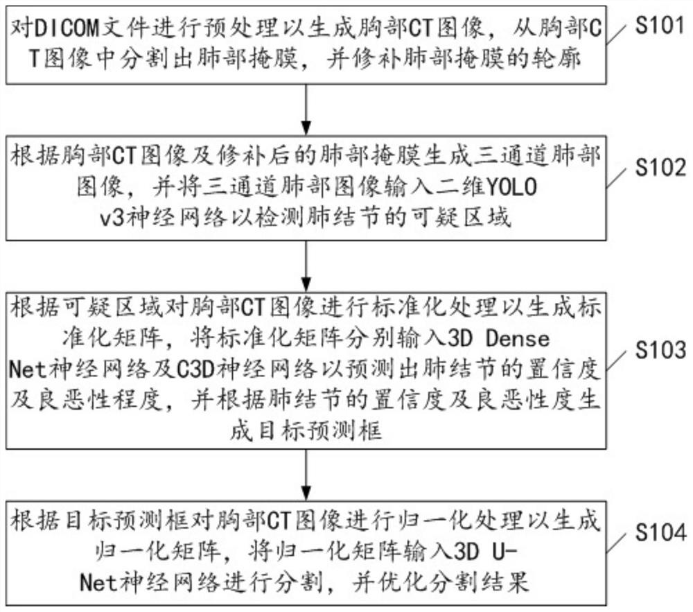
[0035] see figure 1 , figure 1 Shows the flow chart of an embodiment of the deep learning-based pulmonary nodule recognition and segmentation method of the present invention, which includes:

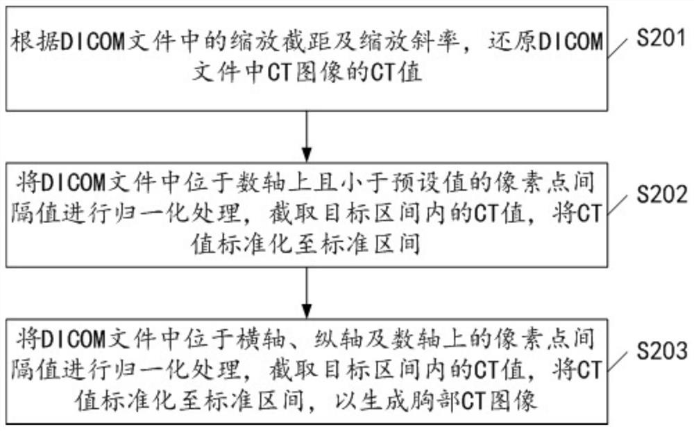
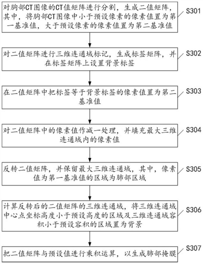
[0036] S101, preprocessing the DICOM file to generate a chest CT image, segmenting a lung mask from the chest CT image, and patching the contour of the lung mask.

[0037] DICOM (Digital Imaging and Communications in Medicine) is an international standard (ISO 12052) for medical images and related information. It defines a medical image format that can be used for data exchange with a quality that meets clinical needs.

[0038] It should be noted that by preprocessing the DICOM file, the real CT value of the CT image in the DICOM file can be effectively restored, and the ...


PUM


Abstract

The invention discloses a pulmonary nodule recognition and segmentation method based on deep learning, and the method comprises the steps: preprocessing a DICOM file so as to generate a chest CT image, segmenting a lung mask from the chest CT image, and repairing the lung mask; generating a three-channel lung image according to the chest CT image and the repaired lung mask, and inputting the three-channel lung image into a two-dimensional YOLO v3 neural network to detect a suspicious area of pulmonary nodules; standardizing the chest CT image according to the suspicious area to generate a standardized matrix, inputting the standardized matrix into a 3D Dense Net neural network and a C3D neural network for prediction, and generating a target prediction box according to a prediction result;and normalizing the chest CT image according to the target prediction box to generate a normalization matrix, inputting the normalization matrix into the 3D UNet neural network for segmentation, and optimizing a segmentation result. The invention further discloses a pulmonary nodule recognition and segmentation system based on deep learning. According to the invention, the accuracy and speed of identification and segmentation can be effectively improved.

Description

technical field [0001] The present invention relates to the technical field of medical image processing, in particular to a method for identifying and segmenting pulmonary nodules based on deep learning and a system for identifying and segmenting pulmonary nodules based on deep learning. Background technique [0002] In the process of identifying pulmonary nodules in images, the commonly used identification methods mainly include the following: [0003] 1. Traditional morphological method: Based on three-dimensional shape pairs, different three-dimensional shapes (such as spherical, cylindrical, and curved surfaces) are used to simulate nodules, blood vessels, and pleura, respectively. However, this method tends to rely on the similarity between the nodule and the spherical shape, and it is easy to cause missed detection due to dissimilarity with the simulated shape. [0004] 2. Radiomics method: On the 3D image, traditional segmentation methods such as OTSU (maximum betwee...

Claims


Application Information

Patent Timeline
IPC IPC(8): G06T7/00G06T7/11G06T7/187G16H30/20G06N3/04
CPCG06T7/0012G06T7/11G06T7/187G16H30/20G06T2207/10081G06T2207/30064G06T2207/20084G06T2207/20081G06N3/045
Inventor 陈海欣李伟忠严朝煜
Owner 广州普世医学科技有限公司
Why Eureka
Try Eureka