
Brain CT image classification method fusing multi-scale superpixels

A CT image and image fusion technology, applied in the field of medical image research, can solve problems such as ignoring the visual characteristics of brain CT images

Pending Publication Date: 2021-04-09
BEIJING UNIV OF TECH
View PDF0 Cites 3 Cited by
  • Summary
  • Abstract
  • Description
  • Claims
  • Application Information

AI Technical Summary

Problems solved by technology

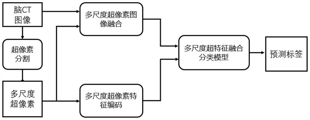
[0005] Aiming at the problem that the above existing methods ignore the visual characteristics of brain CT images, the present invention proposes a brain CT image classification method that fuses multi-scale superpixel fusion (MSF)

Method used


Image

  • Brain CT image classification method fusing multi-scale superpixels
  • Brain CT image classification method fusing multi-scale superpixels
  • Brain CT image classification method fusing multi-scale superpixels

Examples


Embodiment Construction

[0039] In this embodiment, patients with cerebral hemorrhage are used as research objects, but the method is not limited thereto, and brain CT images of patients with other brain diseases can also be used as research objects. Taking the real cerebral hemorrhage CT data set as an example, the implementation steps of this method are described in detail below:

[0040] Step (1) Get data and preprocess:

[0041] Step (1.1) data: the present invention uses the CQ500 data set (http: / / headctstudy.qure.ai / dataset) to collect brain CT images to construct a data set, and actually obtains 451 cases of scan data, a total of 22,773 brain CT images, each patient The label information contains 14 diagnostic categories of brain diseases: intracranial hemorrhage, cerebral parenchymal hemorrhage, ventricular hemorrhage, subdural hemorrhage, epidural hemorrhage, subarachnoid hemorrhage, left cerebral hemorrhage, right cerebral hemorrhage, chronic hemorrhage, Fractures, skull fractures, other fr...


PUM


Abstract

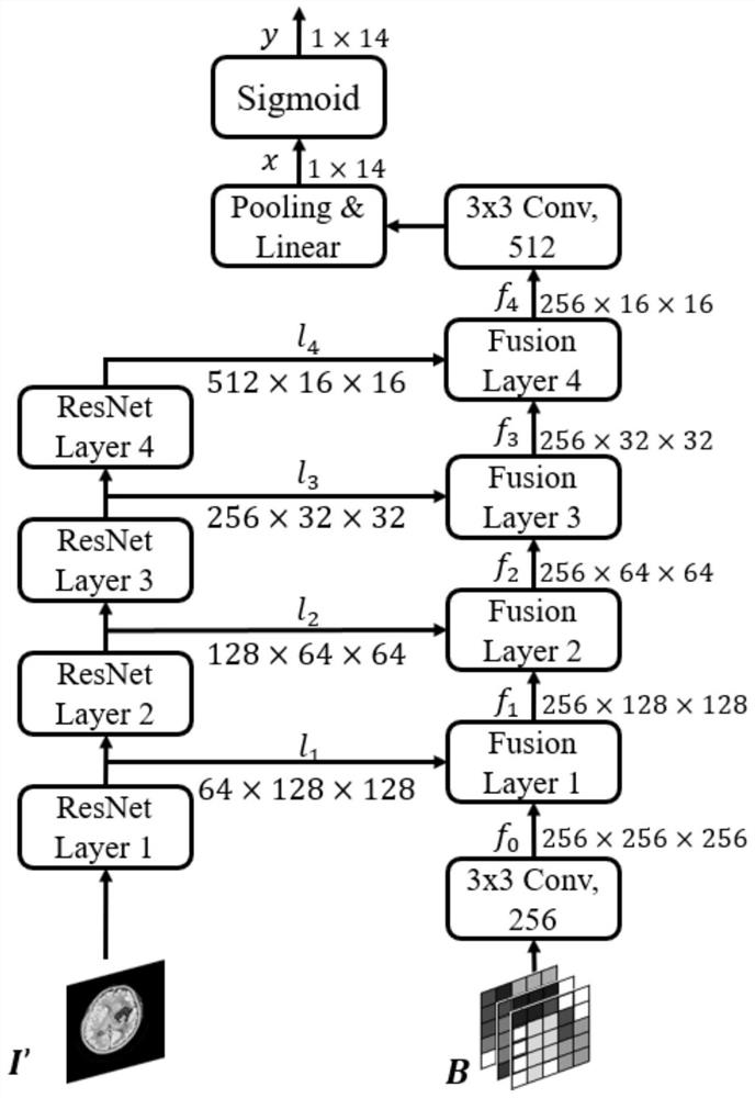
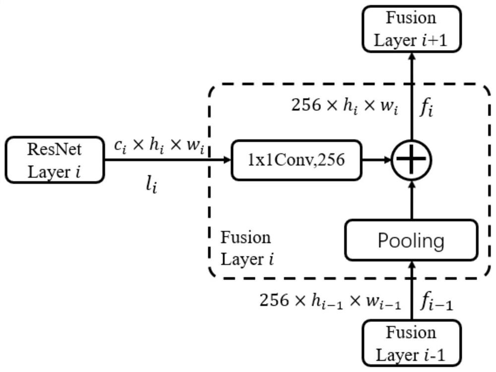
The invention discloses a brain CT image classification method fusing multi-scale superpixels, and belongs to the field of medical image research. The method has the following characteristics: 1) multi-scale superpixels and a brain CT image are fused, image redundant information is removed, and the gray similarity between a focus and surrounding brain tissue pixels is reduced; 2) a multi-scale superpixel encoder based on regions and boundaries is designed, and focus low-level information contained in multi-scale superpixels is effectively extracted; 3) a fusion model fusing multi-scale super-pixel features is designed, and comprehensively utilizing of high-level features extracted by a residual neural network and low-level features of multi-scale super-pixels is carried out to realize classification of brain CT; and 4) compared with the traditional deep learning algorithm, the method provided by the invention can effectively utilize the lesion information contained in the multi-scale superpixels, thereby more accurately classifying the diseases existing in the brain CT image, and the method is reasonable and reliable, and can provide powerful help for the classification of the brain CT image.

Description

technical field [0001] The invention belongs to the field of medical image research, in particular, the invention relates to a method for classifying brain CT images by fusing multi-scale superpixels. Background technique [0002] The diagnosis of brain injury in the clinical emergency department is extremely urgent, and even a short delay can lead to the deterioration of the patient's condition. Computed Tomography (CT) is one of the most commonly used diagnostic tools, which has the characteristics of fast imaging, low cost, wide application range, and high lesion detection rate. Although brain CT can detect critical and time-sensitive abnormalities such as intracranial hemorrhage, increased intracranial pressure, and skull fractures, traditional disease classification methods usually require radiologists to visually assess the size of the hemorrhage area and estimate the midline shift. This process is relatively time-consuming. In recent years, with the advancement and ...

Claims


Application Information

Patent Timeline
IPC IPC(8): G06K9/62G06N3/04G06N3/08G06T5/50G06T7/10
CPCG06N3/08G06T5/50G06T7/10G06T2207/10081G06N3/045G06F18/2453G06F18/253
Inventor 冀俊忠张梦隆张晓丹
Owner BEIJING UNIV OF TECH
Learn More