
Human tissue ultrasonic modeling method, ultrasonic equipment and storage medium

A technology of human tissue and modeling method, which is applied in ultrasonic/sonic/infrasonic diagnosis, ultrasonic/sonic/infrasonic Permian technology, ultrasonic/sonic/infrasonic image/data processing, etc. It can solve difficult problems such as treatment

Pending Publication Date: 2021-05-28
CHISON MEDICAL TECH CO LTD
View PDF0 Cites 0 Cited by
  • Summary
  • Abstract
  • Description
  • Claims
  • Application Information

AI Technical Summary

Problems solved by technology

[0002] Traditional medical imaging devices only provide two-dimensional images of the inside of the human body. Doctors can only estimate the size and shape of the lesion from multiple two-dimensional images based on experience, so as to imagine the three-dimensional geometric relationship between the lesion and its surrounding tissues, which brings great benefits to the treatment. Difficulty

Method used


Image

  • Human tissue ultrasonic modeling method, ultrasonic equipment and storage medium
  • Human tissue ultrasonic modeling method, ultrasonic equipment and storage medium

Examples


Embodiment Construction

[0032] In order to make the purpose, technical solutions and advantages of the embodiments of the present invention clearer, the technical solutions in the embodiments of the present invention will be clearly and completely described below in conjunction with the drawings in the embodiments of the present invention. Obviously, the described embodiments It is a part of embodiments of the present invention, but not all embodiments. Based on the embodiments of the present invention, all other embodiments obtained by those skilled in the art without making creative efforts belong to the protection scope of the present invention.

[0033] Traditional medical imaging devices only provide two-dimensional images of the inside of the human body. Doctors can only estimate the size and shape of the lesion from multiple two-dimensional images based on experience, so as to imagine the three-dimensional geometric relationship between the lesion and its surrounding tissues, which brings great...


PUM


Abstract

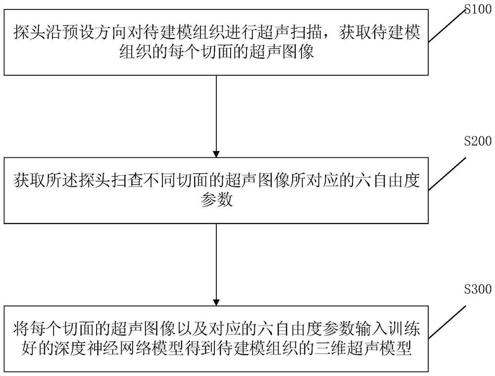
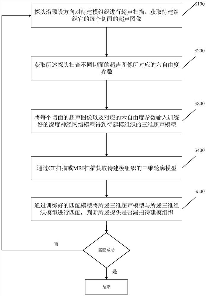
The invention relates to the technical field of image processing, in particular to a human tissue ultrasonic modeling method, ultrasonic equipment and a storage medium. The human tissue ultrasonic modeling method comprises the steps of enabling a probe to conduct ultrasonic scanning on a to-be-modeled tissue in the preset direction to acquire an ultrasonic image of each section of the to-be-modeled tissue; acquiring six-degree-of-freedom parameters corresponding to ultrasonic images of different sections scanned by the probe; and inputting the ultrasonic image of each tangent plane and the corresponding six-degree-of-freedom parameters into a trained deep neural network model to obtain a three-dimensional ultrasonic model of the to-be-modeled tissue. The ultrasound images of the sections of the to-be-modeled tissue can be reconstructed through the trained deep neural network model, the reconstruction accuracy is high, and the efficiency of medical staff for searching the standard section is improved.

Description

technical field [0001] The invention relates to the technical field of ultrasonic image processing, in particular to a human tissue ultrasonic modeling method, ultrasonic equipment and a storage medium. Background technique [0002] Traditional medical imaging devices only provide two-dimensional images of the inside of the human body. Doctors can only estimate the size and shape of the lesion from multiple two-dimensional images based on experience, so as to imagine the three-dimensional geometric relationship between the lesion and its surrounding tissues, which brings great benefits to the treatment. difficult. The 3D visualization technology can reconstruct a 3D shape from a series of 2D images and display it on the terminal. Therefore, not only can the overall concept of the intuitive and image of the imaging object be obtained, but also a lot of important three-dimensional information can be preserved. In addition, because ultrasound imaging has obvious advantages su...

Claims


Application Information

Patent Timeline
Patent Type & Authority Applications(China)
IPC IPC(8): G06T17/00G06T7/73G06T7/00G06N3/08G06N3/04A61B8/08
CPCG06T17/00G06T7/0012G06T7/73G06N3/08A61B8/085G06T2207/10132G06N3/045
Inventor 莫若理
Owner CHISON MEDICAL TECH CO LTD
Learn More