
Patient follow-up analysis

A follow-up, patient technology, applied in the field of patient follow-up analysis, which can solve problems such as restricted access rights and huge data storage capacity

Pending Publication Date: 2021-05-28
SIEMENS HEALTHCARE GMBH
View PDF4 Cites 0 Cited by
  • Summary
  • Abstract
  • Description
  • Claims
  • Application Information

AI Technical Summary

Problems solved by technology

Here, data security and privacy guidelines often severely limit cloud service providers' access to on-premises databases storing medical information
In turn, archiving complete image datasets on cloud platforms for subsequent read-out is generally not a sustainable option as this would require enormous data storage capacity and likewise raises important patient data privacy concerns

Method used


Image

  • Patient follow-up analysis
  • Patient follow-up analysis
  • Patient follow-up analysis

Examples


Embodiment Construction

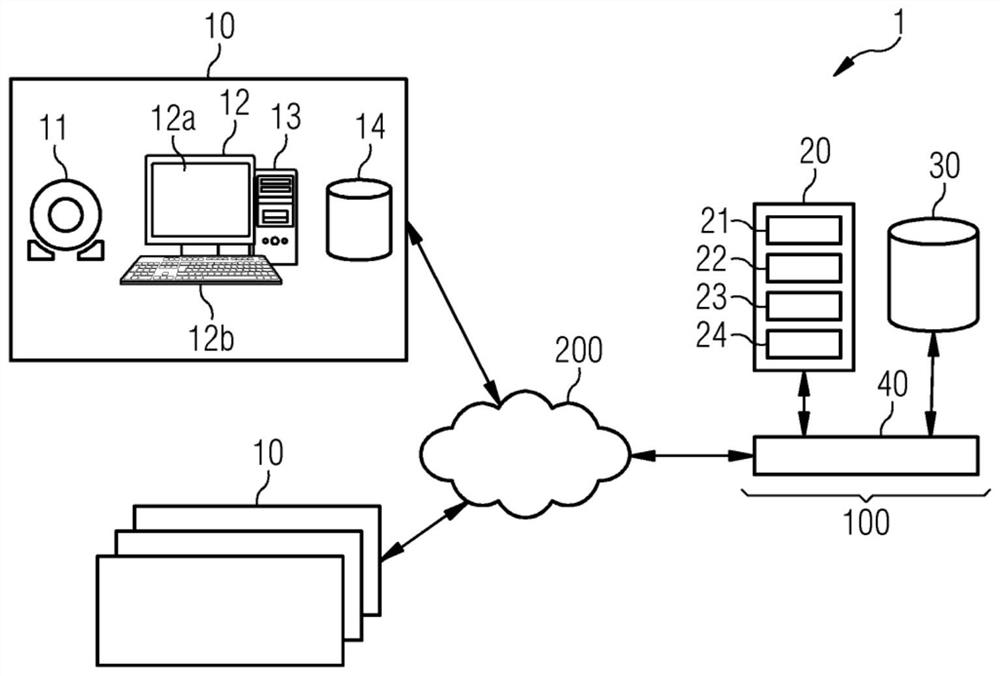
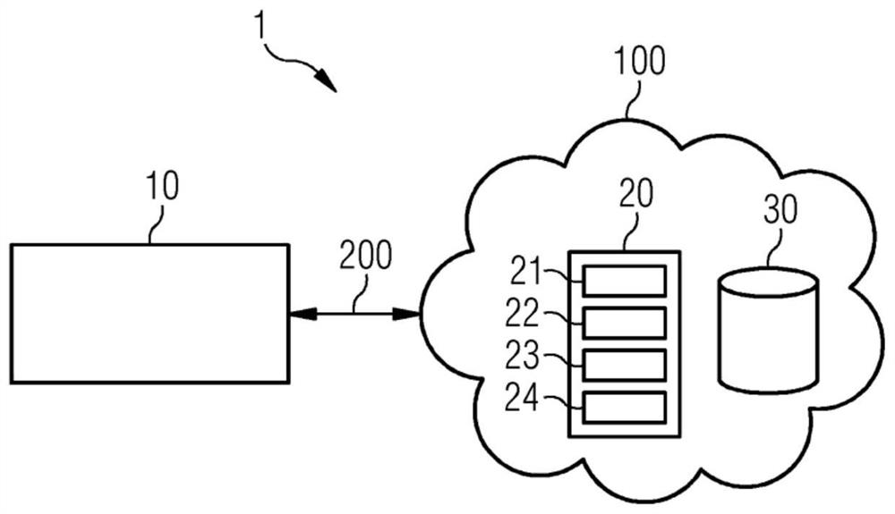
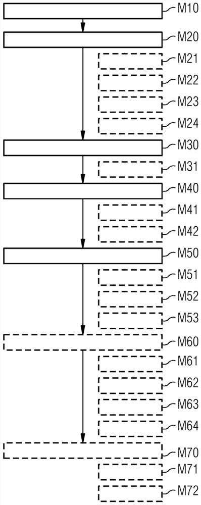
[0089] figure 1 A distributed environment 1 for identifying and quantifying changes in subsequent medical examinations according to an embodiment of the invention is depicted. Environment 1 is suitable for carrying out the method according to one or more embodiments, for example as referenced image 3 and Figure 4 method described further.

[0090] System 1 includes one or more clients 10 and a central processing system 100 . The client 10 and the central processing system 100 are connected via a network 200 . The central processing system 100 is generally configured to control and perform comparisons of data derived from subsequent medical image data in a distributed environment 1 . Client 10 may reside on one or more different local sites. A local site may, for example, relate to a clinical or medical environment such as a hospital or hospital group, laboratory, medical imaging center, clinic or institution. In this example, four clients 10 are shown for illustration....


PUM


Abstract

The invention discloses patient follow-up analysis. A computer-implemented method for comparing follow-up medical findings is provided. The method comprises the steps of receiving image data showing a body part of a patient at a first time and retrieving, from a database, reference data associated to the image data. The reference data comprises reference biometric data and one or more reference medical findings. Thereby the reference biometric data and the one or more reference medical findings have been extracted from reference image data depicting the body part of the patient at a second time different than the first time. The method further comprises processing the received image data so as to extract, from the image data, biometric data corresponding to the reference biometric data and one or more medical findings. Further, the method comprises performing a registration of the extracted biometric data with the reference biometric data. That followed, the extracted medical findings are compared with the reference medical findings using the registration.

Description

technical field [0001] Embodiments relate to subsequent analysis of patient data, such as medical image data. Background technique [0002] Follow-up readings and evaluation of changes are very important when making medical diagnoses from medical images acquired by medical image acquisition modalities such as X-ray equipment, ultrasound equipment, CT or MR scanners. Radiologists often rely not only on the latest available images but on the entire historical record when they make their decisions. The history may include multiple sets of medical image data acquired on the same patient (for the same body part of the patient) at different times and typically using different medical imaging techniques. By tracking such follow-up examinations, changes in medical findings over time can be determined, yielding valuable insights for arriving at correct medical diagnoses. The changes found can for example indicate whether the tumor is growing or the stenosis is getting worse and imm...

Claims


Application Information

Patent Timeline
Patent Type & Authority Applications(China)
IPC IPC(8): G16H10/60G16H50/20G06F16/583
CPCG16H10/60G16H50/20G06F16/583G06T7/0016G06T7/337G06T2207/30048G06T2207/30061G06T7/38G16H30/20G16H30/40G06F21/6245G06T2207/10072
Inventor 乔纳森·斯佩尔斯文·鲍尔卢茨·京德尔雷纳·克格尔
Owner SIEMENS HEALTHCARE GMBH
Learn More