Depth perception enhancement method and device based on 2D/3D blood vessel fusion

A technology of depth perception and blood vessels, applied in the field of medical image processing, can solve problems such as rough blood vessel volume data and unsuitable surfaces

Pending Publication Date: 2021-06-15
ARIEMEDI MEDICAL SCI BEIJING CO LTD
View PDF0 Cites 0 Cited by
  • Summary
  • Abstract
  • Description
  • Claims
  • Application Information

AI Technical Summary

Problems solved by technology

Contour information can help us identify the shape and direction of blood vessels, but the gradient-based

Method used


Image

  • Depth perception enhancement method and device based on 2D/3D blood vessel fusion
  • Depth perception enhancement method and device based on 2D/3D blood vessel fusion
  • Depth perception enhancement method and device based on 2D/3D blood vessel fusion

Examples


Embodiment Construction

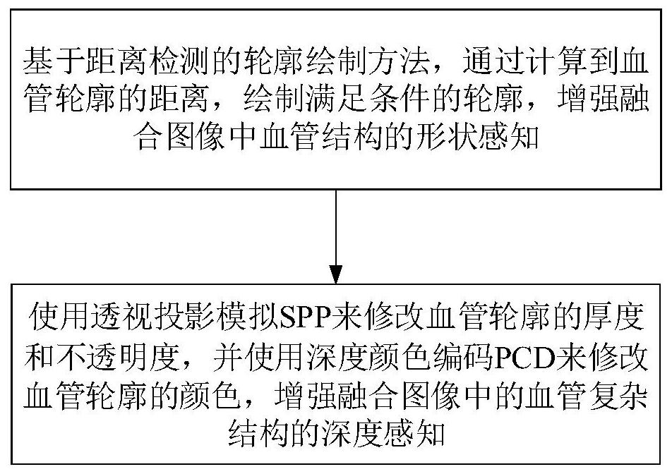
[0017] Such as figure 1 As shown, this depth perception enhancement method based on 2D / 3D blood vessel fusion includes the following steps:

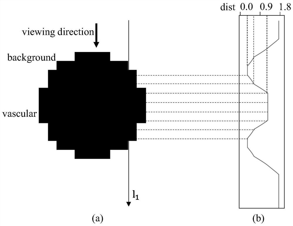
[0018] (1) The contour drawing method based on distance detection, by calculating the distance to the vessel contour, draws the contour satisfying the condition, and enhances the shape perception of the vessel structure in the fusion image;

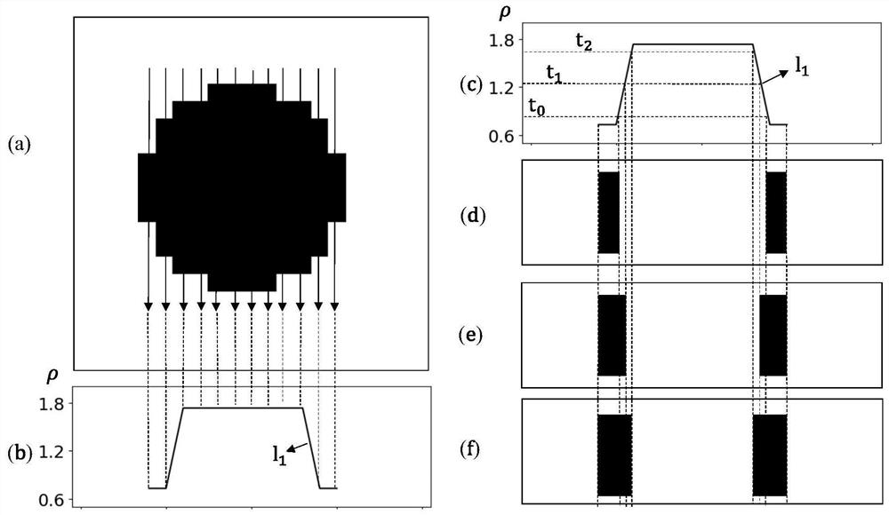
[0019] (2) Use perspective projection to simulate SPP to modify the thickness and opacity of vessel outlines, and use depth color-coded PCD to modify the color of vessel outlines, enhancing the depth perception of complex vessel structures in fused images.

[0020] The contour drawing method based on distance detection in the present invention calculates the distance to the blood vessel contour, draws the contour satisfying the conditions, enhances the shape perception of the blood vessel structure in the fusion image, and then uses perspective projection to simulate SPP to modify the thickness and ...


PUM


Abstract

According to the depth perception enhancement method and device based on 2D/3D blood vessel fusion, depth perception of a blood vessel structure in a fusion image can be enhanced, the method and device are suitable for blood vessel volume data with unsmooth surfaces obtained through CTA segmentation, and shape perception of the blood vessel structure is effectively enhanced. The method comprises the following steps: (1) a contour drawing method based on distance detection draws a contour meeting conditions by calculating the distance to a blood vessel contour, and shape perception of a blood vessel structure in a fused image is enhanced; and (2) modifying the thickness and opacity of the blood vessel contour by using perspective projection simulation SPP, and modifying the color of the blood vessel contour by using depth color coding PCD to enhance the depth perception of the blood vessel complex structure in the fused image.

Description

technical field [0001] The present invention relates to the technical field of medical image processing, in particular to a depth perception enhancement method based on 2D / 3D blood vessel fusion and a depth perception enhancement device based on 2D / 3D blood vessel fusion. Background technique [0002] X-ray fluoroscopy imaging technology can realize intraoperative high spatial and temporal resolution imaging, and has become the current gold standard for diagnosis of clinical vascular interventional surgery (vascular interventional surgery). However, X-ray images are perspective projection imaging. Due to the lack of depth information of vascular structures, doctors cannot fully understand complex three-dimensional vascular anatomical structures such as high curvature and multiple branches. In X-ray image-guided vascular intervention, 2D / 3D image fusion technology will be obtained from preoperative computed tomography angiography (CTA) or magnetic resonance angiography (magne...

Claims


Application Information

Patent Timeline
IPC IPC(8): G06T5/00G06T5/50G06T7/12G06T7/62
CPCG06T5/007G06T5/50G06T2207/10081G06T2207/20221G06T2207/30101G06T7/12G06T7/62
Inventor 杨峰
Owner ARIEMEDI MEDICAL SCI BEIJING CO LTD
Why Eureka
Try Eureka