Magnetic resonance imaging method and device and storage medium

A magnetic resonance imaging and storage medium technology, which is applied in the fields of measuring magnetic variables, medical science, measuring devices, etc., can solve the problems of long magnetic resonance imaging scanning time, and achieve the effect of shortening the magnetic resonance scanning time.

Active Publication Date: 2021-06-25
SHENZHEN CHILDRENS HOSPITAL
View PDF7 Cites 2 Cited by
  • Summary
  • Abstract
  • Description
  • Claims
  • Application Information

AI Technical Summary

Problems solved by technology

[0004] Aiming at the above defects of the prior art, a magnetic resonance imaging method, device and storage me...

Method used


Image

  • Magnetic resonance imaging method and device and storage medium
  • Magnetic resonance imaging method and device and storage medium
  • Magnetic resonance imaging method and device and storage medium

Examples


Embodiment 1

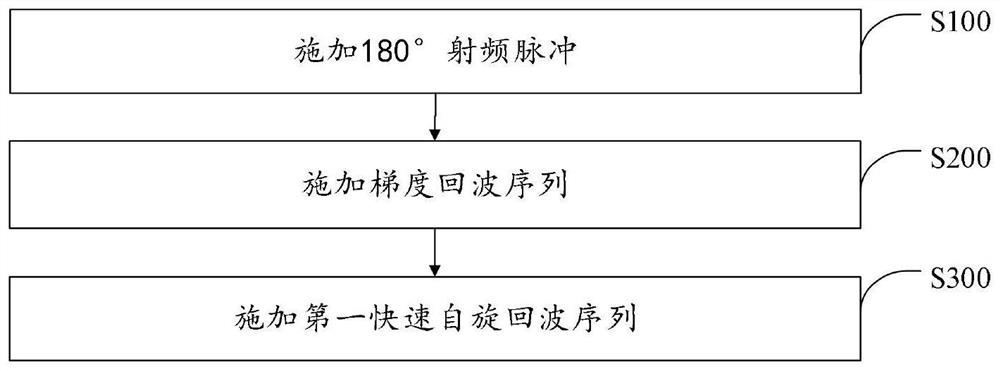
[0041] The magnetic resonance imaging method provided by the present invention applies imaging sequences periodically, and the number of repetitions of the period is not less than 1, such as figure 1 As shown, in each cycle, steps are included:

[0042] S100, applying a 180° radio frequency pulse;

[0043] S200, applying a gradient echo sequence;

[0044] S300. Apply a first fast spin echo sequence.

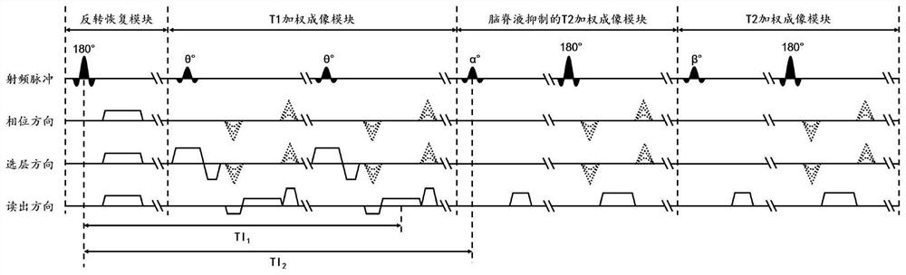
[0045] A T1-weighted image is generated from the data acquired in the gradient echo sequence, and a CSF-suppressed T2-weighted image is generated from the data acquired in the first fast spin echo sequence. Specifically, such as figure 2 As shown, the magnetic resonance imaging equipment periodically applies an imaging sequence, and each cycle of the imaging sequence includes a reversal recovery module, a T1-weighted imaging module, and a T2-weighted module for cerebrospinal fluid suppression. The sequence diagram of each module is shown in Figure 4-6 As shown, the sequence...

Embodiment 2

[0060] Based on the above embodiments, the present invention also provides a magnetic resonance imaging device, the block diagram of which can be as follows Figure 8 shown. The device includes a processor 10 and a memory 20 . Understandably, Figure 8 Only some components of the terminal are shown, but it should be understood that implementation of all illustrated components is not required, and more or fewer components may be implemented instead.

[0061] The storage 20 may be an internal storage unit of the terminal in some embodiments, such as a hard disk or memory of the terminal. In other embodiments, the memory 20 may also be an external storage device of the terminal, such as a plug-in hard disk equipped on the terminal, a smart memory card (Smart Media Card, SMC), a secure digital (Secure Digital, SD ) card, flash memory card (Flash Card), etc. Further, the memory 20 may also include both an internal storage unit of the terminal and an external storage device. Th...

Embodiment 3

[0064] The present invention also provides a storage medium, where one or more programs are stored in the storage medium, and the one or more programs can be executed by one or more processors, so as to realize the steps of the magnetic resonance imaging method described in the above-mentioned embodiments .


PUM


Abstract

The invention discloses a magnetic resonance imaging method and device and a storage medium. The method comprises the following steps of: in each period, applying 180-degree radio frequency pulse, applying a gradient echo sequence, and applying a first fast spin echo sequence, the number of repetition times of the period being not less than 1; and generating a T1 weighted image according to the data acquired by the gradient echo sequence, and generating a cerebrospinal fluid suppressed T2 weighted image according to the data acquired by the first fast spin echo sequence. According to the method, the gradient echo sequence is applied after the 180-degree radio frequency pulse is applied and before the first fast spin echo sequence is applied, T1 weighted image data is acquired by utilizing idle time between a turnover recovery pulse and an excitation pulse required for generating the cerebrospinal fluid suppressed T2 weighted image, and the magnetic resonance scanning time for generating the T1 weighted image and the cerebrospinal fluid suppressed T2 weighted image is shortened.

Description

technical field [0001] The present invention relates to the field of magnetic resonance technology, in particular to a magnetic resonance imaging method, equipment and storage medium. Background technique [0002] Clinically, magnetic resonance sequences are often used to scan and image lesions. For some diseases, such as TSC (Tuberous Sclerosis Complex, Tuberous Sclerosis Complex), a clearer display is required. In order to clearly display lesions, the selected clinical scanning The intra-layer resolution is high, and the amount of data to be collected is large, resulting in a longer scanning time, and a longer scanning time will cause discomfort to the patient. [0003] Therefore, the prior art still needs to be improved and improved. Contents of the invention [0004] In view of the above-mentioned defects of the prior art, a magnetic resonance imaging method, device and storage medium are provided, aiming at solving the problem of long scanning time of the magnetic re...

Claims


Application Information

Patent Timeline
IPC IPC(8): G01R33/561A61B5/055
CPCG01R33/5618G01R33/561A61B5/055
Inventor 胡湛棋廖建湘赵彩蕾赵霞袁碧霞叶园珍操德智朱凤军姚一曾洪武干芸根王海峰苏适杨俊
Owner SHENZHEN CHILDRENS HOSPITAL
Why Eureka
Try Eureka