
Ultrasonic imaging method, ultrasonic imaging equipment and computer storage medium

An ultrasonic imaging method and ultrasonic imaging technology, applied in the directions of ultrasonic/sonic/infrasonic diagnosis, acoustic diagnosis, infrasonic diagnosis, etc.

Pending Publication Date: 2021-07-02
SHENZHEN MINDRAY BIO MEDICAL ELECTRONICS CO LTD
View PDF0 Cites 0 Cited by
  • Summary
  • Abstract
  • Description
  • Claims
  • Application Information

AI Technical Summary

Problems solved by technology

This display method and ultrasound report form have certain limitations. The final diagnosis result given by the doctor is obtained by combining the lesion characteristics of multiple slice images. In general, only one ultrasound image cannot fully reflect the lesion. Lesion features

Method used


Image

  • Ultrasonic imaging method, ultrasonic imaging equipment and computer storage medium
  • Ultrasonic imaging method, ultrasonic imaging equipment and computer storage medium
  • Ultrasonic imaging method, ultrasonic imaging equipment and computer storage medium

Examples


Embodiment Construction

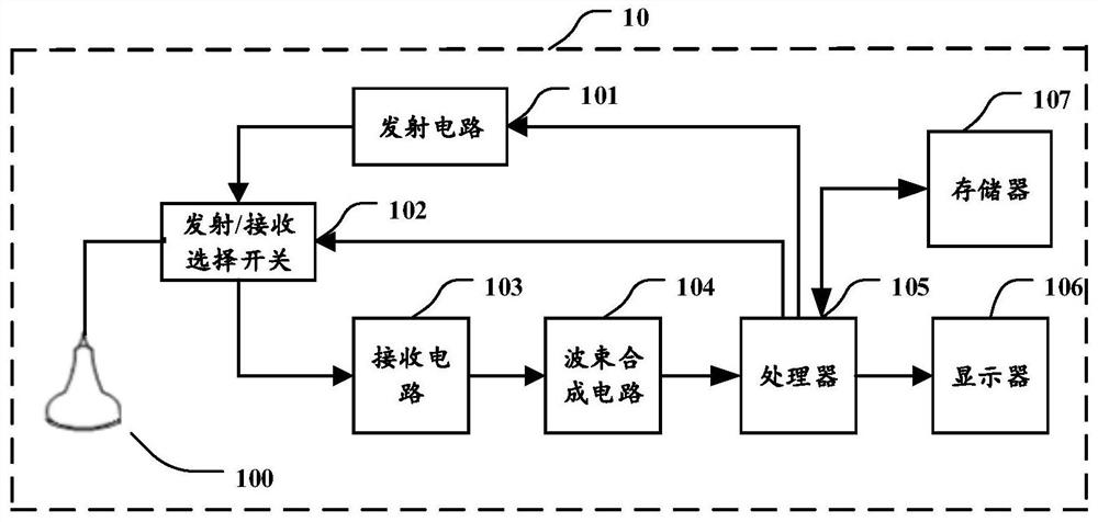
[0030] figure 1 A structural block diagram of an ultrasound imaging system in the present application embodiment. The ultrasonic imaging system 10 can include a probe 100, a transmitting circuit 101, an transmitting / reception selection switch 102, a receiving circuit 103, a beam synthesis circuit 104, a processor 105, and a display 106. The transmitting circuit 101 can excite the probe 100 to emit ultrasonic waves to the tissue to be tested; the receiving circuit 103 can receive ultrasonic echo signal / data returned from the tissue by the probe 100, thereby obtaining an ultrasonic echo signal / data; the ultrasonic echo signal / data passes beamped After the synthesis circuit 104 performs beam synthesis, it is sent to the processor 105. The processor 105 processes the ultrasonic echo signal / data to obtain an ultrasound image to be tissue. The ultrasound image obtained by the processor 105 can be stored in the memory 107. These ultrasound images can be displayed on display 10...


PUM


Abstract

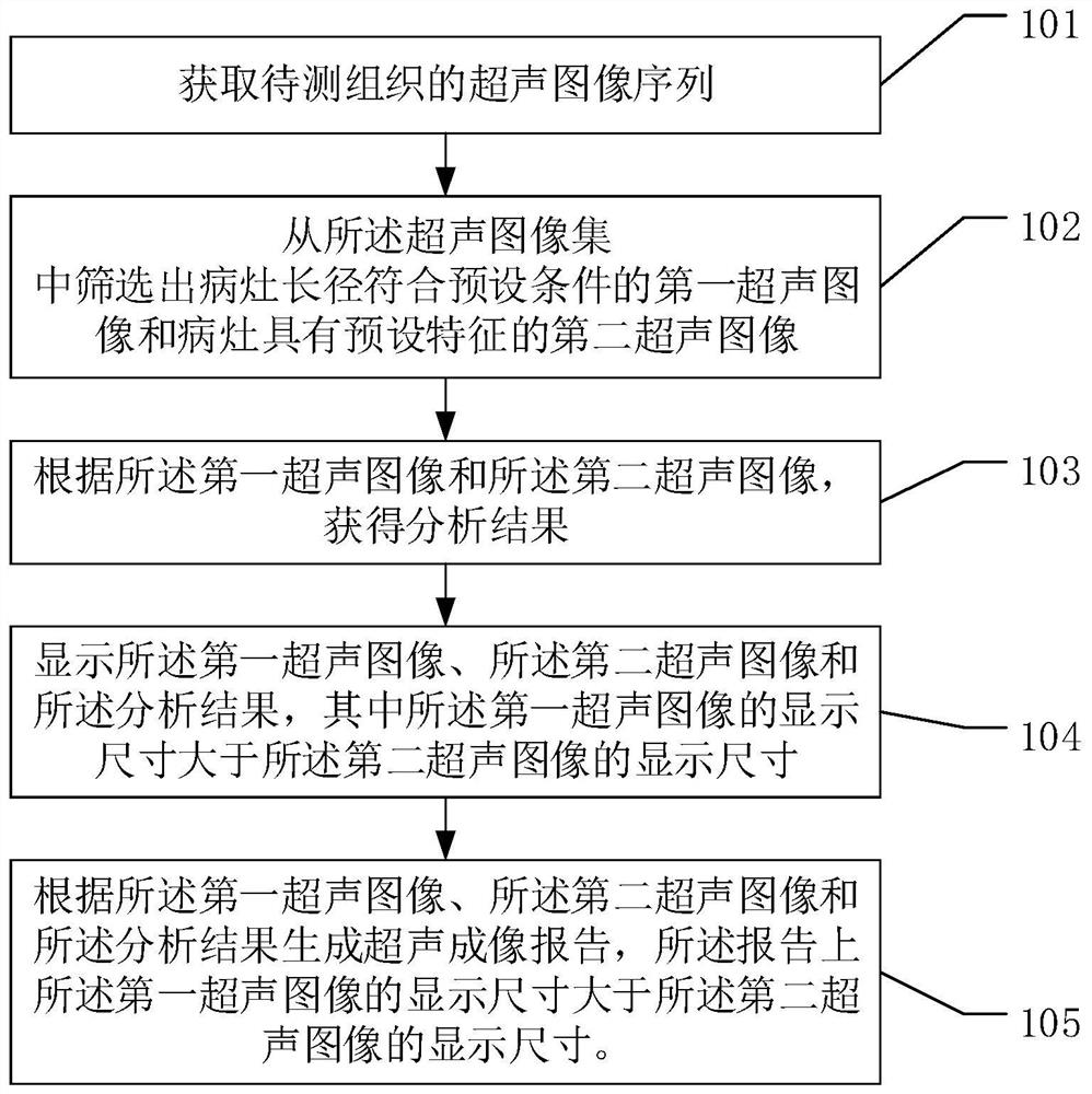
The invention discloses an ultrasonic imaging method, ultrasonic imaging equipment and a computer storage medium. The ultrasonic imaging method comprises the following steps: acquiring multiple frames of ultrasonic images of to-be-detected tissue; screening out a first ultrasonic image in which the long diameter of a lesion meets a preset condition and a second ultrasonic image in which the lesion has a preset feature from the multiple frames of ultrasonic images; acquiring an analysis result; displaying the first ultrasonic image, the second ultrasonic image and the analysis result, wherein the display size of the first ultrasonic image is larger than the display size of the second ultrasonic image; and generating an ultrasonic imaging report, wherein the display size of the first ultrasonic image on the report is larger than the display size of the second ultrasonic image. According to the invention, the first ultrasonic image and the second ultrasonic image are displayed at the same time, and the display size of the first ultrasonic image is larger than the display size of the second ultrasonic image, so a user can focus on a key ultrasonic image to comprehensively know the focus condition.

Description

Technical field [0001] The present application relates to the field of medical devices, and more particularly to an ultrasound imaging method, an ultrasound imaging apparatus, and a computer storage medium. Background technique [0002] Ultrasonic image inspection has become the preferred solution for clinical diagnosis due to non-invasive, simple, low price, repeatable operation. For lesions such as thyroid, mammary gland, clinical diagnosis, doctors typically scan the lesion characteristics of multiple sections of the lesions to obtain a more accurate diagnosis result. [0003] Currently, only one ultrasound image is displayed on the ultrasound imaging device, only one section diagram with lesions is displayed on the ultrasound report and use text descriptions. Such display modes and ultrasound report have certain limitations, and the final diagnosis result given the diagnosis of the doctor is obtained by the lesionic characteristics of the integrated multi-cut surface, and in ...

Claims


Application Information

Patent Timeline
Patent Type & Authority Applications(China)
IPC IPC(8): A61B8/00
CPCA61B8/463A61B8/5223A61B8/5238A61B8/5292
Inventor 李若平丛龙飞安兴
Owner SHENZHEN MINDRAY BIO MEDICAL ELECTRONICS CO LTD
Learn More