

Kit and method for extracting DNA of FFPE tissue sample and application thereof
A technology of tissue samples and kits, which is applied in the field of DNA extraction kits for FFPE tissue samples, can solve the problems of low DNA extraction, long dewaxing time, and low purity, so as to reduce manual operation procedures, shorten dewaxing time, The effect of high purity
- Summary
- Abstract
- Description
- Claims
- Application Information
AI Technical Summary
Problems solved by technology
Method used
Image

Examples
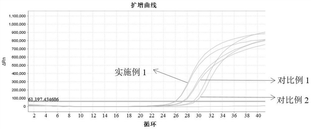
Embodiment 1
[0064] 1. Scrape FFPE tissue samples (5-10μm thick, 1×1cm 2 size) 1-4 pieces into a 1.5mL centrifuge tube, add 300μL deparaffinization buffer (2% SDS, 200mM Tris-HCl, 10mM EDTA-2Na, 1% Triton X-100, 0.1M NaCl , 1% Tween 20, 2% PEG6000), and then add 20 μL of proteinase K to shake and mix, and incubate in a metal bath at 55° C. for 15 minutes.
[0065] 2. Raise the temperature of the metal bath to 80°C and continue to incubate for 15 minutes (note: it is not necessary to take out the centrifuge tube, the temperature can be directly raised from 55°C to 80°C). The phase solution (about 250 μL) was aspirated and transferred to a new 1.5mL centrifuge tube.
[0066] 3. Add 30 μL of magnetic beads and 300 μL of binding solution (65% isopropanol, 0.2M NaCl) to the centrifuge tube in turn, mix by pipetting, and then mix every 2 minutes for 3 times to avoid magnetic Beads gather.
[0067] 4. Place the centrifuge tube on the magnetic stand for 30 seconds, so that the magnetic beads ar...
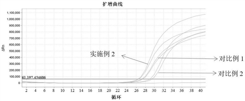
Embodiment 2
[0074] 1. Scrape FFPE tissue samples (5-10μm thick, 1×1cm 2 size) 1-4 pieces into a 1.5mL centrifuge tube, add 300μL deparaffinization buffer (3% SDS, 100mM Tris-HCl, 5mM EDTA-2Na, 0.5% Triton X-100, 0.2M NaCl , 0.1% Tween 20 and 0.1% PEG6000), 20 μL of proteinase K, mixed by shaking, and incubated at 56° C. for 15 min.
[0075] 2. Raise the temperature of the metal bath to 80°C, and continue to incubate for 15 minutes (Note: It is not necessary to take out the centrifuge tube, the temperature can be directly raised from 55°C to 80°C). The aqueous phase solution (about 250 μL) was aspirated and transferred to a new 1.5mL centrifuge tube.
[0076] 3. Add 20 μL of magnetic beads and 300 μL of binding solution (80% isopropanol and 0.4M NaCl) in sequence, shake and mix, and then mix every 3 minutes for 3 times to avoid aggregation of magnetic beads.
[0077] 4. Place the centrifuge tube on the magnetic stand for 30 seconds, so that the magnetic beads are completely absorbed. Ca...
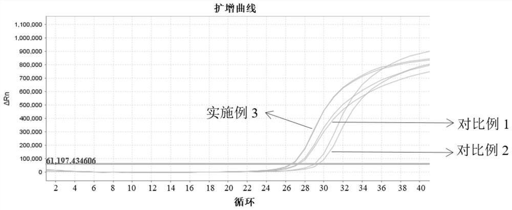
Embodiment 3
[0084] 1. Scrape FFPE tissue samples (5-10μm thick, 1×1cm 2 size) 1-4 pieces into a 1.5mL centrifuge tube, add 300μL deparaffinization buffer (2% SDS, 200mM Tris-HCl, 10mM EDTA-2Na, 1% Triton X-100, 0.1M NaCl, 1% Tween 20, 2% PEG6000), and then add 20 μL of proteinase K to shake and mix, and incubate in a metal bath at 55° C. for 30 min.
[0085] 2. Raise the temperature of the metal bath to 80°C and continue to incubate for 30 minutes (Note: It is not necessary to take out the centrifuge tube, the temperature can be raised directly from 55°C to 80°C). The aqueous solution was aspirated and transferred to a new 1.5mL centrifuge tube.
[0086] Subsequent steps are the same as in Example 1.
PUM
 Abstract
Description
Claims
Application Information