Arterial intracavity ultrasonic image processing method and related device

A technology of intracavity ultrasound and image processing, which is applied in the directions of ultrasound/sonic/infrasonic image/data processing, ultrasound/sonic/infrasonic diagnosis, ultrasound/sonic/infrasonic Permian technology, etc. It can solve problems that cannot be intuitive and real Reflects the severity of diseased tissue and the inaccurate measurement of long-axis images

Active Publication Date: 2021-07-16
SONOSCAPE MEDICAL CORP
View PDF5 Cites 2 Cited by
  • Summary
  • Abstract
  • Description
  • Claims
  • Application Information

AI Technical Summary

Problems solved by technology

[0006] The purpose of this application is to provide a method, device, equipment, readable storage medium, and ultrasonic diagnostic system for image processing in the arterial cavity, aiming to solve the problem of being unintuitive and realistic due to the fact that it still performs equal-pitch helical motion by default during image playback. It reflects the severity of the diseased tissue, and the long-axis image directly generated during the acquisition process has the problem of inaccurate measurement

Method used


Image

  • Arterial intracavity ultrasonic image processing method and related device
  • Arterial intracavity ultrasonic image processing method and related device
  • Arterial intracavity ultrasonic image processing method and related device

Examples


Embodiment 1

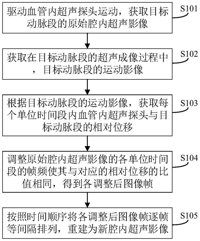
[0081] See figure 1 , figure 1 A flow chart of a method for processing intraarterial ultrasound images provided in an embodiment of the present application, which includes the following steps:

[0082] S101: Drive the movement of the intravascular ultrasound probe to obtain the original intracavity ultrasound image of the target artery segment;

[0083] This step aims to obtain the original intracavity ultrasound image of the target artery segment collected by the intravascular ultrasound probe by driving the intravascular ultrasound probe to move in the arterial cavity by the intravascular ultrasound image processing device. It should be understood that the acquisition frequency of the original intraarterial ultrasound images is a unified default frequency, that is, each unit time period has the same frame rate, which can be called the default frame rate. Usually, the default frame rate It can be 30FPS, 60FPS or even higher 120FPS (it means that the ultrasound image with a ...

Embodiment 2

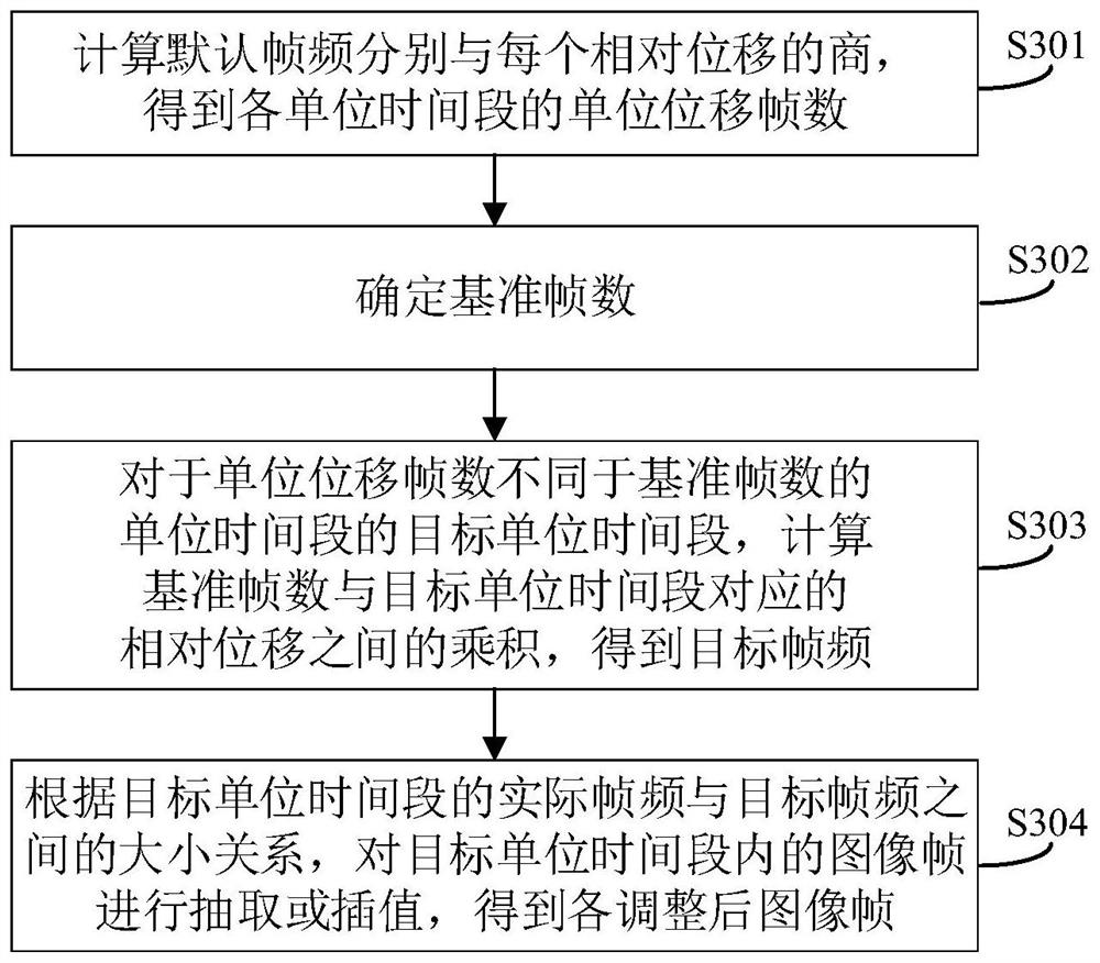
[0107] Since the ratio of the frame frequency to its corresponding relative displacement can be expressed as the number of frames per unit displacement, the adjustment of the frame frequency can also be converted to the adjustment of the number of frames per unit displacement, that is, it is only necessary to adjust to the same number of frames per unit displacement. This embodiment provides a image 3 The feasible implementation scheme shown in the flowchart includes the following steps:

[0108] S301: Calculate the quotient of the default frame rate and each relative displacement to obtain the number of frames per unit displacement in each unit time period;

[0109] This step aims to use the default frame rate as the numerator, each relative displacement as the denominator, and calculate the number of frames per unit displacement by division, which represents the number of frames corresponding to each unit displacement of the intravascular ultrasound probe.

[0110] For eas...


PUM


Abstract

The invention discloses an arterial intracavity ultrasonic image processing method. The method comprises the following steps of acquiring relative displacement between an intravascular ultrasonic probe and a target artery section within each unit time period according to a motion image of a target artery section, and taking the relative displacement as a basis for guiding the adjustment of the frame frequency within each unit time period, i.e., making targeted adjustment to same original frame frequency within each unit time period of the original intracavity ultrasonic image: adjusting the frame frequency within each unit time of ultrasonic images in an original cavity to enable the ratio of the frame frequencies to the corresponding relative displacement to be the same. In this way, new arterial intracavity images obtained through reconstruction on the adjusted frame images can visually and truly reflect the severity degree of the pathological tissue when replayed, and meanwhile, the situation of inaccurate measurement of long-axis images directly generated in the collection process is avoided. The invention further discloses an arterial intracavity ultrasonic image processing device and equipment, a readable storage medium and an ultrasonic diagnosis system, all of which have the foregoing beneficial effects.

Description

technical field [0001] The present application relates to the technical field of ultrasonic image processing, in particular to a method, device, equipment, readable storage medium, and ultrasonic diagnostic system for intraarterial ultrasonic image processing. Background technique [0002] In the process of using the probe to go deep into the coronary cavity to acquire intracoronary images, the probe will make a variable pitch in the intraluminal tissue due to the influence of cardiac contraction, relaxation, and the expansion and retraction of the vessel lumen with blood pressure changes. The spiral motion, that is, the precession length of the spiral motion per unit time is different, which is reflected in the collected coronary intracavity images, that is, the corresponding displacement difference between adjacent frame images is different. [0003] During the examination of intracoronary artery disease, the doctor's diagnosis of the diseased tissue and the determination ...

Claims


Application Information

Patent Timeline
IPC IPC(8): A61B8/12A61B8/08
CPCA61B8/12A61B8/0891A61B8/4245
Inventor 洪杰韩黎英云张勇朱彦聪
Owner SONOSCAPE MEDICAL CORP
Why Eureka
Try Eureka