
Medical image classification method and device, computer equipment and storage medium

A medical image and classification method technology, applied in the field of image recognition, can solve the problems of reduced performance and generalization, high similarity, model deviation, etc., and achieve the effect of improving learning ability, enhancing feature extraction ability, and avoiding frequent changes

Active Publication Date: 2021-07-16
CHANGSHA UNIVERSITY OF SCIENCE AND TECHNOLOGY
View PDF5 Cites 5 Cited by
  • Summary
  • Abstract
  • Description
  • Claims
  • Application Information

AI Technical Summary

Problems solved by technology

[0004] Existing medical image recognition algorithms cannot overcome problems such as information loss, gradient disappearance, and degradation when the network is transmitted as the number of network layers deepens. At the same time, the similarity between categories of colon polyp images is high and the variability within categories is low. , can lead to model bias and overfitting, leading to poor performance and generalization

Method used


Image

  • Medical image classification method and device, computer equipment and storage medium
  • Medical image classification method and device, computer equipment and storage medium
  • Medical image classification method and device, computer equipment and storage medium

Examples


Embodiment Construction

[0027] In order to make the purpose, technical solution and advantages of the present application clearer, the present application will be further described in detail below in conjunction with the accompanying drawings and embodiments. It should be understood that the specific embodiments described here are only used to explain the present application, and are not intended to limit the present application.

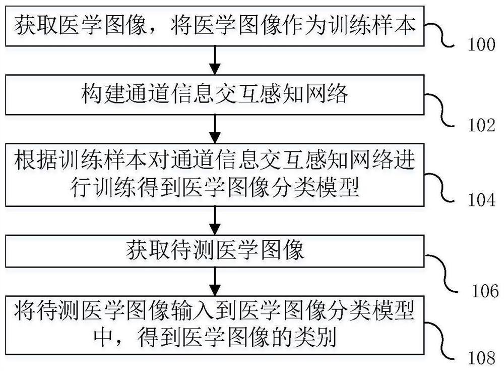
[0028] In one embodiment, such as figure 1 As shown, a medical image classification method is provided, the method includes the following steps:

[0029] Step 100, acquire medical images, and use the medical images as training samples.

[0030] The colonoscopy images taken by the olympus PCF-H290DI equipment were randomly selected from the database of the gastrointestinal endoscopy room of a certain hospital. Before labeling, the colonoscopy images were first cropped, and the white edges around them were removed. The size of the images was unified to 256*256, and then Ha...


PUM


Abstract

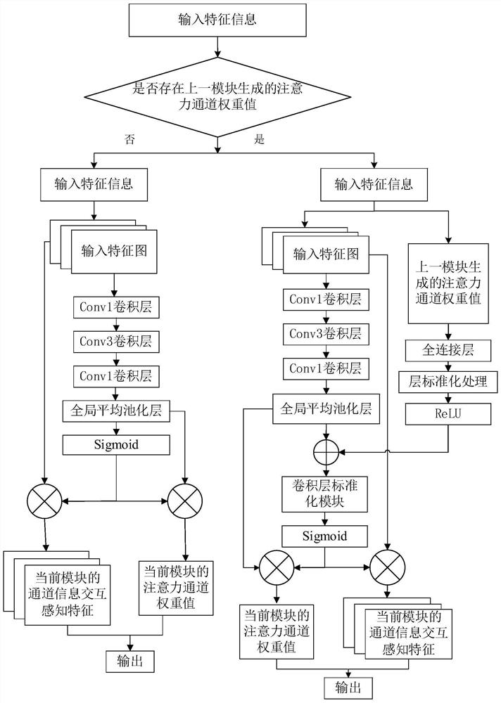
The invention relates to a medical image classification method and device, computer equipment and a storage medium, and the method comprises the steps: obtaining a medical image, and taking the medical image as a training sample; constructing a channel information interaction perception network, wherein the network comprises an input network, a feature extraction network and an output network; training the channel information interaction perception network according to the training sample to obtain a medical image classification model; and acquiring a to-be-tested medical image, and inputting the to-be-tested medical image into the medical image classification model to obtain the category of the medical image. According to the method, the feature of ta previous module and the currently extracted feature are combined, so that it is guaranteed that information flows among the modules in a feed-forward mode, frequent change of the information among the modules is effectively avoided, the learning ability of the modules is improved, and the feature extraction ability of the network is enhanced; and after the pathological picture of a patient is transmitted to the trained network model, a diagnosis result can be directly given, so that the working efficiency of a doctor can be improved, the doctor is effectively helped to reduce the missed diagnosis rate, and the diagnosis accuracy is improved.

Description

technical field [0001] The present application relates to the technical field of image recognition, in particular to a medical image classification method, device, computer equipment and storage medium. Background technique [0002] Currently, the common clinical methods for the detection of colorectal cancer include fecal occult blood test, optical colonoscopy, and sigmoidoscopy. Among them, the biopsy of polyp tissue through optical colonoscopy to determine whether there is tumor, the nature of the tumor and the degree of differentiation is the gold standard for colon examination. However, if all the polyps found are removed and pathological biopsy is performed, a large amount of labor costs will be invested, which is not conducive to vigorously promoting the prevention and research of colorectal cancer. In order to improve the detection rate of precancerous polyps, in addition to improving the inspection level of endoscopists through systematic training and rich practica...

Claims


Application Information

Patent Timeline
Patent Type & Authority Applications(China)
IPC IPC(8): G06T7/00G06K9/62G06N3/04G06N3/08G16H50/20
CPCG06T7/0012G06N3/084G16H50/20G06T2207/10068G06T2207/30028G06T2207/30096G06N3/045G06F18/214
Inventor 王威胡意晖王新李骥周思远
Owner CHANGSHA UNIVERSITY OF SCIENCE AND TECHNOLOGY
Learn More