Sheep fetal skin fibroblast and separation method and application thereof

A fibroblast and fibroblast technology, applied in the field of cell engineering, can solve the problems of less neutralizing antibodies, less passage of primary cells, and small intra-batch production volume, etc., to accelerate cell metabolism, promote cell growth and reproduction, morphology uniform effect

Active Publication Date: 2021-08-06
LANZHOU INST OF VETERINARY SCI CHINESE ACAD OF AGRI SCI
View PDF9 Cites 0 Cited by
  • Summary
  • Abstract
  • Description
  • Claims
  • Application Information

AI Technical Summary

Problems solved by technology

[0003] The preparation of vaccines for the prevention of oral ulcers mainly includes the following aspects: first, through continuous separation and cultivation of primary cells of cattle or sheep, the oral oral ulcer virus is isolated from them, and then continuously passaged for attenuation to prepare attenuated vaccines; second, the current There is also a trend of research and development of inactivated vaccines; third, because the surface of the sheep oral ulcer virus is covered with a capsule shaped like a small tube, which is arranged in a criss-cross pattern to cover its epitope, resulting in less neutralizing antibodies produced in the sheep, and the protection rate for sheep In view of the particularity of this immunology, some researchers used ultrasound and other means to destroy the capsule of ORFV and then prepare a mouth sore vaccine. However, the above situations are limited to laboratory research and have not been used in production large scale use
The main reasons are: the separation of primary cells is time-consuming, laborious and expensive, the number of passages of primary cells is small, the toxicity of the cultured aphthus virus is low, the production volume within a batch is small, and the repeatability between batches is difficult to be consistent due to differences in primary cells

Method used


Image

  • Sheep fetal skin fibroblast and separation method and application thereof
  • Sheep fetal skin fibroblast and separation method and application thereof
  • Sheep fetal skin fibroblast and separation method and application thereof

Examples


Embodiment 1

[0031] Separation and optimization of sheep fetal skin fibroblasts

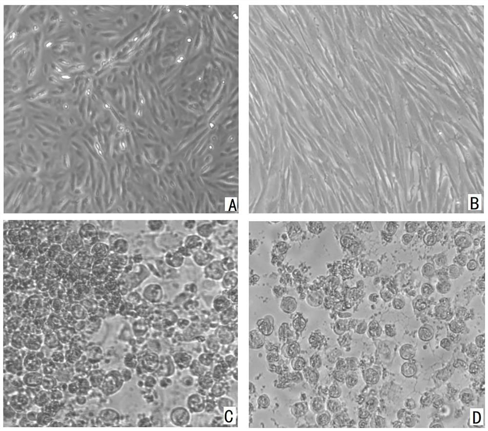
[0032] Routinely aseptically take out the sheep fetus, cut a small piece of testicular skin tissue, wash it several times with PBS with a pH of 7.0 and a concentration of 0.05mol / L, cut it into pieces aseptically, and then use type IV collagenase (4mg / ml) Digest for 0.5-1h, add 2ml of fetal bovine serum, centrifuge at 1500rpm for 10min, discard the supernatant, resuspend the pellet in digestion solution containing 0.05% trypsin and 0.02%EDTA, incubate at 37°C for 10-30min, add 5% fetal bovine serum Stop the digestion, then filter with one layer, two layers of high-pressure sterilized gauze, 100 mesh and 200 mesh filters in sequence. During the filtration period, the filtrate is continuously diluted with PBS, and the final filtrate is centrifuged at 1500rpm for 10min, and the precipitate is sheep fetal skin Primary fibroblast cells were resuspended or frozen in DMEM / F12 containing 10% fetal bovine serum.

[0...

Embodiment 2

[0037] Tolerance of SFSFS cells to different concentrations of h-EGF

[0038] Since the cells need 10% fetal bovine serum to grow well, reducing the concentration of fetal bovine serum will affect the cell growth. However, fetal bovine serum is expensive and difficult to use for industrial production of ORFV vaccines.

[0039] In Example 2, on the basis of Example 1, a medium that is non-toxic to SFSFS cells and can reduce the amount of fetal bovine serum used is screened.

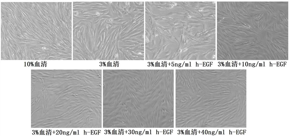
[0040] Divide SFSFS cells at 1.0x10 6 The concentration of each / ml was divided into 3% fetal bovine serum+5ng / ml h-EGF, 3% fetal bovine serum+10ng / ml h-EGF, 3% fetal bovine serum+20ng / ml h-EGF, 3 % fetal bovine serum+30ng / ml h-EGF, 3% fetal bovine serum+40ng / ml h-EGF in DMEM / F12 medium, and cells containing 10% and 3% fetal bovine serum medium were set as controls. Cultivate for 48h, observe the cell growth situation, the results are as follows: image 3 shown.

[0041] image 3 The results showed th...

Embodiment 3

[0043] CCK-8 detection results at different time points after different concentrations of h-EGF acted on SFSFS cells

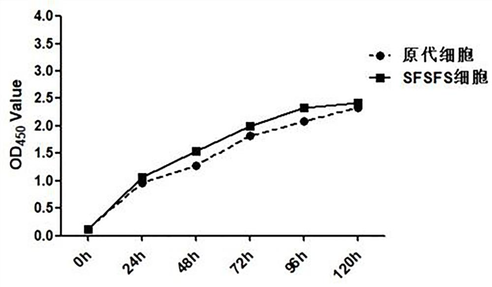
[0044] In order to clarify the effect of different concentrations of h-EGF on the viability of SFSFS cells at different time points, 10% fetal bovine serum, 3% fetal bovine serum, 3% fetal bovine serum + 5ng / ml h-EGF, 3% fetal bovine serum Bovine serum + 10ng / ml h-EGF, 3% fetal bovine serum + 20ng / ml h-EGF, 3% fetal bovine serum + 30ng / ml h-EGF, 3% fetal bovine serum + 40ng / ml h -EGF and DMEM / F12 were used to form the medium, and the cells were divided into 96-well plates, and five replicate wells were set up for each medium, and the wells without cells were set as negative controls. At different time points, after adding CCK-8 for 2 hours, measure OD 450 values, cell growth curves and toxicity test results such as figure 2 and Figure 4 shown.

[0045] Figure 4 The results showed that at 24 hours, the SFSFS cells cultured with 10% fetal bovine serum we...


PUM


Abstract

The invention discloses a sheep fetal skin fibroblast and a separation method and application thereof. The preservation number of the fibroblast is CCTCC No: C2019202. In the separation process of the sheep fetal skin fibroblast SFSFS, digestive juice containing 0.05% trypsin and 0.02% EDTA and IV-type collagenase are adopted for treating sheep fetal skin tissue, and then digestive juice containing 0.01-0.05% trypsin-EDTA is used for treating cells; and 3% fetal calf serum and 10-40ng / ml h-EGF are adopted for replacing 10% fetal calf serum to culture the sheep fetal skin fibroblast, the use amount of the fetal calf serum is greatly reduced, the SFSFS culture cost and the aphtha virus production cost are reduced, and resources are saved. The cell strain can obviously increase the separation rate of aphtha viruses and shorten the separation time, the separated aphtha virus titer and virus copy number are higher, and a tool can be provided for large-scale reproduction of the aphtha viruses and virus infection pathogenesis research.

Description

technical field [0001] The invention relates to the technical field of cell engineering, in particular to a sheep fetal skin fibroblast, and also relates to a separation method and application of the cell. Background technique [0002] Contagious Ethyma (CE), commonly known as sheep mouth sore, is an acute and contagious infectious disease caused by sheep mouth ulcer virus (ORFvirus, ORFV) to which goats, sheep, and mainly lambs are susceptible. Zoonotic diseases that can infect humans and other animals. ORFV is mainly prevalent in Inner Mongolia, Tibet, Xinjiang, Yunnan, Gansu, Sichuan, Ningxia and other sheep-intensive areas. In recent years, it has frequently occurred in Ningxia, Inner Mongolia, Xinjiang, Tibet, Shandong, Hubei, Yunnan, Shaanxi, Heilongjiang, Qinghai and other regions . The typical symptoms of cauliflower-like hyperplasia will appear on the lips, oral mucosa, tongue and nostrils of affected sheep, which seriously affects their feeding and production, an...

Claims


Application Information

Patent Timeline
IPC IPC(8): C12N5/073C12N5/071C12N7/00C12N7/02C12R1/91
CPCC12N5/0625C12N5/0603C12N7/00C12N2509/00C12N2509/10C12N2501/11C12N2710/24251Y02A50/30
Inventor 吴锦艳尚佑军杜国玉候俊玲尚振华刘丹李玲霞康赛红
Owner LANZHOU INST OF VETERINARY SCI CHINESE ACAD OF AGRI SCI
Why Eureka
Try Eureka