Craniocerebral CT angiography image processing method and device, medium and electronic device

An angiography and image technology, applied in the field of image processing, can solve the problems of difficulty in ensuring accuracy, inability to quickly and accurately determine the state of the brain region, and spending a lot of time to resolve, so as to ensure segmentation accuracy and avoid anatomical structures. Unreasonable, the effect of improving segmentation efficiency

Pending Publication Date: 2021-09-24
沈阳东软智能医疗科技研究院有限公司
View PDF0 Cites 1 Cited by
  • Summary
  • Abstract
  • Description
  • Claims
  • Application Information

AI Technical Summary

Problems solved by technology

At this stage, the segmental blood supply areas are mainly distinguished by the naked eye. Usually, only experienced doctors can quickly and accurately identify the segmental blood supply areas with the naked eye, while inexperienced doctors need to spend a lot of time to distinguish the segmental blood supply areas, and the accuracy is relatively low. Difficult to guarantee, in this way, it will not be possible to quickly and accurately determine the state of the brain region

Method used


Image

  • Craniocerebral CT angiography image processing method and device, medium and electronic device
  • Craniocerebral CT angiography image processing method and device, medium and electronic device
  • Craniocerebral CT angiography image processing method and device, medium and electronic device

Examples


Embodiment Construction

[0056] Specific embodiments of the present disclosure will be described in detail below in conjunction with the accompanying drawings. It should be understood that the specific embodiments described here are only used to illustrate and explain the present disclosure, and are not intended to limit the present disclosure.

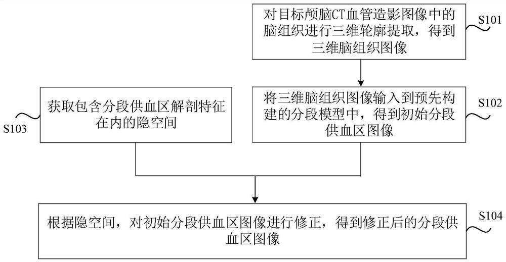
[0057] As discussed in the background technology, in order to determine the status of brain regions, doctors usually need to visually distinguish multiple segmental blood supply areas in brain CTA images, but only experienced doctors can quickly and accurately distinguish each segment with the naked eye blood supply area, and inexperienced doctors need to spend a lot of time to distinguish each segment of blood supply area, and the accuracy is difficult to guarantee. For this reason, it is necessary to use a computer to automatically divide multiple segmental blood supply areas in the CTA image. At present, it mainly depends on the accurate registration of the...


PUM


Abstract

The invention relates to a craniocerebral CT angiography image processing method and device, a medium and an electronic device. The method comprises the following steps: performing three-dimensional contour extraction on brain tissues in a target craniocerebral CT angiography image to obtain a three-dimensional brain tissue image; inputting the three-dimensional brain tissue image into a pre-constructed segmented model to obtain an initial segmented blood supply area image; acquiring a hidden space including the anatomical features of the segmented blood supply area; and correcting the initial segmented blood supply area image according to the hidden space. In this way, the multiple segmented blood supply areas can be automatically obtained only by inputting the three-dimensional brain tissue image corresponding to the target craniocerebral CT angiography image into the segmentation model, and the segmentation efficiency is remarkably improved. Besides, the hidden space including the anatomical features of the segmented blood supply area is fused into the correction process of the segmentation result, so that the problem that the segmentation result is unreasonable in the aspect of the anatomical structure due to the fact that the segmentation model cannot learn the anatomical features of the segmented blood supply area in the training stage can be avoided, and the segmentation precision is guaranteed.

Description

technical field [0001] The present disclosure relates to the technical field of image processing, and in particular, relates to a processing method, device, medium and electronic equipment for brain CT angiography images. Background technique [0002] CT angiography (Computed Tomography angiography, CTA) image refers to the image obtained by CT scanning under the condition of injecting contrast agent into human blood vessels, and the blood vessels are more obvious. Compared with computed tomography without contrast agent, CTA can clearly display the vertebral artery, basilar artery, posterior cerebral artery and its main branches, and can provide an important basis for diagnosis of occlusive vascular lesions. The relationship with blood vessels is of great value. [0003] Among them, brain CTA images usually include multiple segmental blood supply areas. Doctors can determine the status of brain regions by analyzing the CTA image features of each segmental blood supply area...

Claims


Application Information

Patent Timeline
Patent Type & Authority Applications(China)
IPC IPC(8): G06T7/00G06T7/12G06T7/13G06T7/181G06T5/00G06K9/62G06K9/46
CPCG06T7/0012G06T7/12G06T7/13G06T7/181G06T5/002G06T2207/10012G06T2207/10081G06T2207/20032G06T2207/20081G06T2207/30101G06F18/241
Inventor 杨明雷钱山袁红美贾晓甜
Owner 沈阳东软智能医疗科技研究院有限公司
Why Eureka
Try Eureka