Eureka AIR delivers breakthrough ideas for toughest innovation challenges, trusted by R&D personnel around the world.

Eyelid tumor digital pathological section image multi-classification method based on deep learning

A technology for pathological sectioning and eyelid tumors, applied in image analysis, image enhancement, image data processing, etc., to achieve high accuracy

Active Publication Date: 2021-09-28
ZHEJIANG UNIV
View PDF5 Cites 3 Cited by
  • Summary
  • Abstract
  • Description
  • Claims
  • Application Information

AI Technical Summary

Problems solved by technology

However, the application of AI in the field of pathology still faces great challenges because the format of histopathology images is difficult to be read by neural networks.

Method used


Image

  • Eyelid tumor digital pathological section image multi-classification method based on deep learning
  • Eyelid tumor digital pathological section image multi-classification method based on deep learning
  • Eyelid tumor digital pathological section image multi-classification method based on deep learning

Examples


Embodiment Construction

[0060] The present invention will be further described below in conjunction with the accompanying drawings and specific embodiments.

[0061] Embodiments of the present invention and its implementation process are as follows:

[0062] The hardware environment used for implementation is: CPU Intel(R), GPU is NVIDIA RTX2080Ti, and the operating environment is Python3.6 and Pyrorch 0.4.1.

[0063] Step 1. Data acquisition:

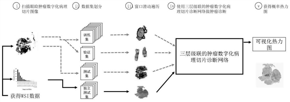
[0064] The pathological slices of eyelid tumors classified by known lesion categories are scanned to obtain digital pathological slice images of eyelid tumors, and a training set is constructed from all digital pathological slice images of eyelid tumors; figure 1 As shown, in the specific implementation, a data set can be constructed from all digital pathological slice images of eyelid tumors, and then the data set can be divided into training set, verification set, and test set.

[0065] Step 2, data augmentation:

[0066] Aiming at the problems of uneven...


PUM


Abstract

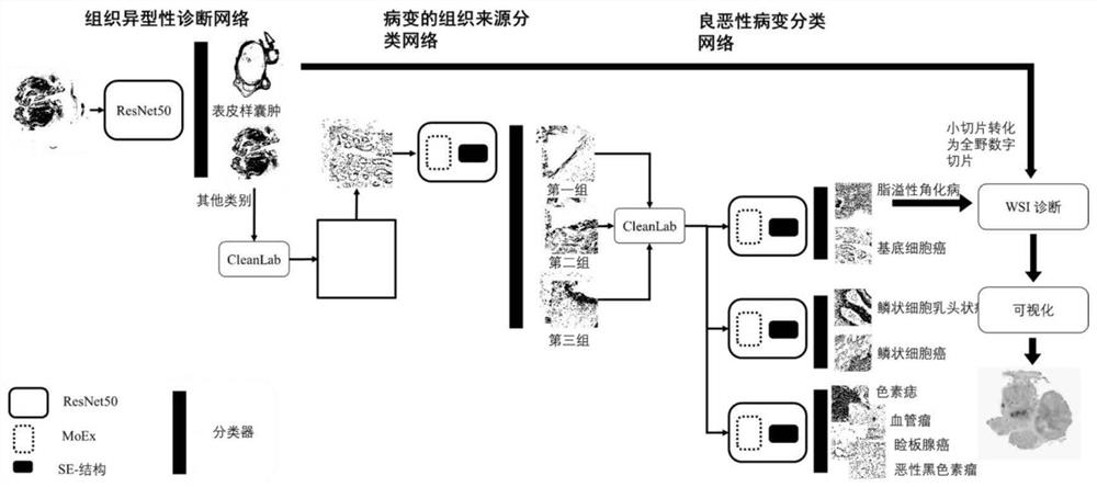
The invention discloses an eyelid tumor digital pathological section image multi-classification method based on deep learning. The method comprises steps of scanning eyelid tumor pathological sections classified by known lesion categories to obtain an image construction training set; performing data enhancement and normalization processing, constructing a three-layer cascaded tumor digital pathological section diagnosis network, training the tumor digital pathological section diagnosis network by using an enhanced training set, performing prediction processing, generating a probability thermodynamic diagram, and performing lesion category detection. The method can effectively visualize the position and lesion type of the tumor in the full-field digital slice to assist in diagnosis, perform preliminary screening and prompting of lesion areas, change the attention of the network to channels and improve the network performance.

Description

technical field [0001] The present invention relates to a multi-classification method for eyelid images in the field of deep learning, computer vision and ophthalmic tumors, in particular to a multi-classification method for digitized pathological slice images of eyelid tumors based on deep learning, which uses deep learning technology to digitize eyelid tumors A method for detecting and analyzing pathological slice images. Background technique [0002] Computer vision is a kind of artificial intelligence technology, which refers to the use of computers to simulate human vision, which is the "seeing" in artificial intelligence. In terms of technical process, it is divided into three parts: target detection, target recognition, and behavior recognition. According to the target type of recognition, it can be divided into image recognition, object recognition, face recognition, text recognition and so on. In the field of intelligent robots, computer vision can perform feature...

Claims


Application Information

Patent Timeline
IPC IPC(8): G06K9/62G06T7/00G16H50/20
CPCG06T7/0012G16H50/20G06T2207/30041G06T2207/30096G06F18/2411G06F18/253
Inventor 叶娟王琳艳吕岱霖王亚奇孙玲玲邵安金凯
Owner ZHEJIANG UNIV
Why Eureka
Eureka Blog
Learn More