
Image enhancement equipment based on animal tissue slice imaging

A technology of animal tissue and image enhancement, which is applied in image enhancement, image data processing, and analysis of solids using sound waves/ultrasonic waves/infrasonic waves, etc. It can solve the problems of inaccurate control of the scanning mechanism and differences in detection results, and achieve detection stability and detection Comprehensive improvement, good detection effect, and improved control accuracy

Active Publication Date: 2021-11-16
QIQIHAR MEDICAL UNIVERSITY
View PDF6 Cites 2 Cited by
  • Summary
  • Abstract
  • Description
  • Claims
  • Application Information

AI Technical Summary

Problems solved by technology

[0005] To this end, the present invention provides an image enhancement device based on animal tissue slice imaging, which is used to overcome the inaccurate control of the scanning mechanism during the scanning process of the image enhancement equipment based on animal tissue slice imaging in the prior art. difference problem

Method used


Image

  • Image enhancement equipment based on animal tissue slice imaging
  • Image enhancement equipment based on animal tissue slice imaging
  • Image enhancement equipment based on animal tissue slice imaging

Examples


Embodiment Construction

[0064] In order to make the objects and advantages of the present invention clearer, the present invention will be further described below in conjunction with the examples; it should be understood that the specific examples described here are only for explaining the present invention, and are not intended to limit the present invention.

[0065] Preferred embodiments of the present invention are described below with reference to the accompanying drawings. Those skilled in the art should understand that these embodiments are only used to explain the technical principle of the present invention, and are not intended to limit the protection scope of the present invention.

[0066] It should be noted that, in the description of the present invention, terms such as "upper", "lower", "left", "right", "inner", "outer" and other indicated directions or positional relationships are based on the terms shown in the accompanying drawings. The direction or positional relationship shown is ...


PUM


Abstract

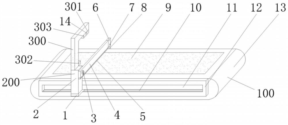
The invention relates to image enhancement equipment based on animal tissue slice imaging, and relates to the technical field of animal tissue imaging. The equipment comprises: a bearing mechanism, wherein the bearing mechanism comprises a bottom plate and a photosensitive plate connected with the bottom plate, and the bearing mechanism is used for bearing the animal tissue slice; a fixing mechanism which is connected with the bearing mechanism, wherein the fixing mechanism comprises a sliding seat connected with one side of a bottom plate of the bearing mechanism, a supporting plate connected with the sliding seat, a rotating shaft connected with the supporting plate, a first motor connected with one end of the rotating shaft and a pressing plate connected with the other end of the rotating shaft, and the fixing mechanism is used for fixing the animal tissue slices; and a scanning mechanism which is connected with the fixing mechanism, wherein the scanning mechanism comprises an ultrasonic line scanner used for detecting the position and flatness of the animal tissue slice and a camera used for detecting the thickness of the animal tissue slice. When in use, the animal tissue slice detection device can better adapt to animal tissue slices with different sizes, so that the detection stability and comprehensiveness of the animal tissue slices are improved, and the accuracy of a detection result is further improved.

Description

technical field [0001] The invention relates to the technical field of animal tissue imaging, in particular to an image enhancement device based on animal tissue slice imaging. Background technique [0002] Animal tissue is the basic structure of multicellular animals composed of cells and intercellular substances with similar morphological functions. It is divided into four types: epithelial tissue, connective tissue, muscle tissue and nervous tissue. They are interconnected and interdependent in different proportions to form animal tissues. Various organs and systems in order to complete various physiological activities. Animal tissues are differentiated from the original inner, middle and outer embryonic layers during the embryonic period; animal tissues can be differentiated according to their origin, morphological structure and function. The common characteristics are divided into four categories: epithelial tissue, connective tissue, muscle tissue and nervous tissue. ...

Claims


Application Information

Patent Timeline
Patent Type & Authority Applications(China)
IPC IPC(8): G01N29/06G01N29/22G01B11/00G01B11/06G01B17/08G01N21/59G06T5/00
CPCG01N29/0654G01N29/22G01N29/223G01B11/00G01B11/06G01B17/08G01N21/59G06T5/00G01N2291/0237G01N2291/0289G01N2291/2632
Inventor 姚宏波梁明辉文丽波宫学武张萌王月静
Owner QIQIHAR MEDICAL UNIVERSITY
Learn More