Medical image multi-organ segmentation method and system

A medical image and multi-organ technology, applied in the field of medical image processing, can solve the problems of different shapes of organs and tissues, different shapes of organs and tissues, and difficulty in obtaining accurate segmentation at the boundary, so as to improve the segmentation performance, improve the accuracy, The effect of avoiding the degradation of segmentation accuracy

Active Publication Date: 2021-12-17
SHANDONG UNIV
View PDF4 Cites 3 Cited by
  • Summary
  • Abstract
  • Description
  • Claims
  • Application Information

AI Technical Summary

Problems solved by technology

[0008] In order to solve the deficiencies of the prior art, the present invention provides a medical image multi-organ segmentation method and system, which solves the problems in the medical image segmentation process that the organs and tissues within the subject and between subjects are different, and different The different shapes of organs and tissues lead to segmentation difficulties, and solve the problem of difficult to obtain accurate segmentation in some areas, especially at the border, caused by the low contrast between tissues and organs and the background environment

Method used


Image

  • Medical image multi-organ segmentation method and system
  • Medical image multi-organ segmentation method and system
  • Medical image multi-organ segmentation method and system

Examples


Embodiment 1

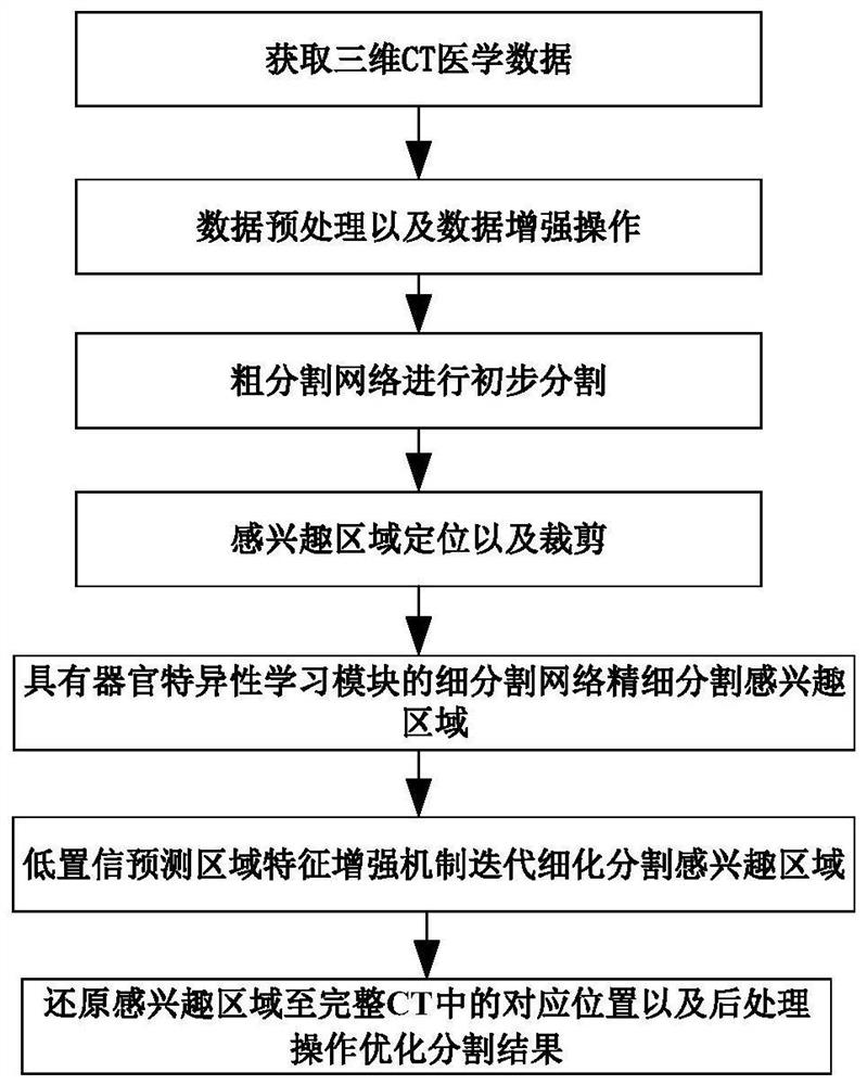
[0057] In one or more implementations, a method for multi-organ segmentation of medical images is disclosed, referring to figure 1 shown, including the following steps:

[0058] Step 1: Obtain the 3D CT medical data to be segmented.

[0059] The step 1 specifically includes:

[0060] Step 1.1: Use the SimpleITK library to read a set of CT image data in nifti compressed format.

[0061] Step 1.2: Use the built-in function GetArrayFromImage provided by the SimpleITK library to convert the SimpleITKImage format into numpy format for subsequent processing.

[0062] Step 2: Perform data preprocessing and data enhancement operations on the acquired 3D CT medical data to be segmented.

[0063] Described step 2 specifically comprises:

[0064] Step 2.1: Adjust the window width and level of the CT image data so as to increase the contrast of the CT image.

[0065] Specifically, according to the task to be processed, the window bottom win_min and window top win_max of the window ra...

Embodiment 2

[0158] In one or more embodiments, a medical image multi-organ segmentation system is disclosed, including the following modules:

[0159] An acquisition module configured to: acquire three-dimensional CT medical data to be segmented, perform specific data preprocessing operations and data enhancement operations on the medical data;

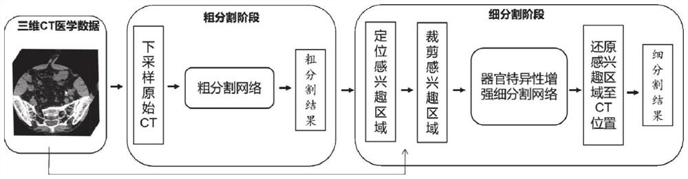
[0160] The rough segmentation module is configured to: down-sampling three-dimensional CT medical data to obtain low-resolution data, and using a rough stage segmentation network to obtain a down-sampled segmentation prediction result;

[0161] The fine segmentation module is configured to: upsample the segmentation prediction result of the rough segmentation module to the original resolution, use the segmentation prediction result of the rough segmentation module to locate the region of interest of the 3D CT medical data, clip the region of interest, and input Based on the organ-specific dynamic adjustment fine-stage segmentation network, the or...

Embodiment 3

[0167] A computer-readable storage medium stores a plurality of instructions, and the instructions are suitable for being loaded by a processor of a terminal device and executing the method for segmenting multiple organs of a medical image provided in Embodiment 1.


PUM


Abstract

The invention discloses a medical image multi-organ segmentation method and system. The method comprises the following steps: carrying out data preprocessing and enhancement operation on acquired to-be-segmented three-dimensional CT medical data; performing down-sampling to obtain low-resolution data, and inputting the low-resolution data into a rough stage segmentation network to obtain a down-sampling segmentation prediction result; performing up-sampling on the down-sampled segmentation prediction result to an original resolution ratio, and positioning an area of interest of the three-dimensional CT medical data by using the segmentation prediction result in the rough stage and cutting out the area of interest; inputting the region of interest into a fine stage segmentation network based on organ specificity dynamic adjustment for fine segmentation; further refining and segmenting by using an iterative refined low-confidence prediction region feature enhancement technology to obtain a segmentation result of the region of interest; processing the obtained segmentation result, the original resolution and the cutting coordinates, restoring the region of interest to the corresponding position in the complete CT, obtaining the final segmentation result through post-processing operation, and the segmentation performance and the segmentation precision are improved.

Description

technical field [0001] The invention relates to the technical field of medical image processing, in particular to a method and system for segmenting multiple organs of a medical image. Background technique [0002] The statements in this section merely set forth background technical information related to the present invention and do not necessarily constitute prior art. [0003] Medical image segmentation refers to the segmentation of pixels / voxels of anatomical or pathological structures of interest from medical images, which is an important step in the process of medical image processing and analysis. As an important part of medical image processing technology, medical image segmentation is often a preprocessing step in clinical applications such as computer-aided diagnosis, radiation therapy planning, computer-aided surgery planning, disease treatment and prognosis, and plays a key role in the field of clinical diagnosis. However, the traditional manual segmentation met...

Claims


Application Information

Patent Timeline
Patent Type & Authority Applications(China)
IPC IPC(8): G06T7/11G06N3/04G06N3/08
CPCG06T7/11G06N3/08G06T2207/10081G06T2207/20081G06T2207/20084G06T2207/20104G06T2207/20132G06T2207/30004G06N3/045
Inventor 周元峰刘尽华
Owner SHANDONG UNIV
Why Eureka
Try Eureka