
Cancerous cell detection method and system

A detection method and cancer cell technology, applied in the field of cancer cell detection method and system, can solve the problems of missing cancer cells and not using results, etc., and achieve the effect of improving detection efficiency and accuracy

Pending Publication Date: 2022-01-04
上海申挚医疗科技有限公司
View PDF0 Cites 0 Cited by
  • Summary
  • Abstract
  • Description
  • Claims
  • Application Information

AI Technical Summary

Problems solved by technology

[0002] At present, the traditional diagnosis is to make glass slides by staining, and the laboratory experts will view them one by one through a microscope. Due to the relatively large field of view of the slides and the large number of nuclei, it takes a long time for experts to determine the diagnosis of a pathological slide. , and there is a high possibility of missing possible cancer cells due to visual fatigue
[0003] In addition, because each examiner has his own subjective judgment, it is easy to cause the same film to be checked by different doctors, and there will be different results.

Method used


Image

  • Cancerous cell detection method and system
  • Cancerous cell detection method and system
  • Cancerous cell detection method and system

Examples


Embodiment Construction

[0060] Preferred embodiments of the present invention will be described in detail below in conjunction with the accompanying drawings.

[0061] In order to further understand the present invention, the preferred embodiments of the present invention are described below in conjunction with examples, but it should be understood that these descriptions are only to further illustrate the features and advantages of the present invention, rather than limiting the claims of the present invention.

[0062] The description in this part is only for several typical embodiments, and the present invention is not limited to the scope of the description of the embodiments. The mutual replacement of the same or similar prior art means and some technical features in the embodiments is also within the scope of the description and protection of the present invention.

[0063] The description of the steps in each embodiment in the specification is only for convenience of description, and the imple...


PUM


Abstract

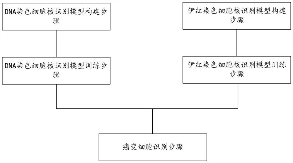
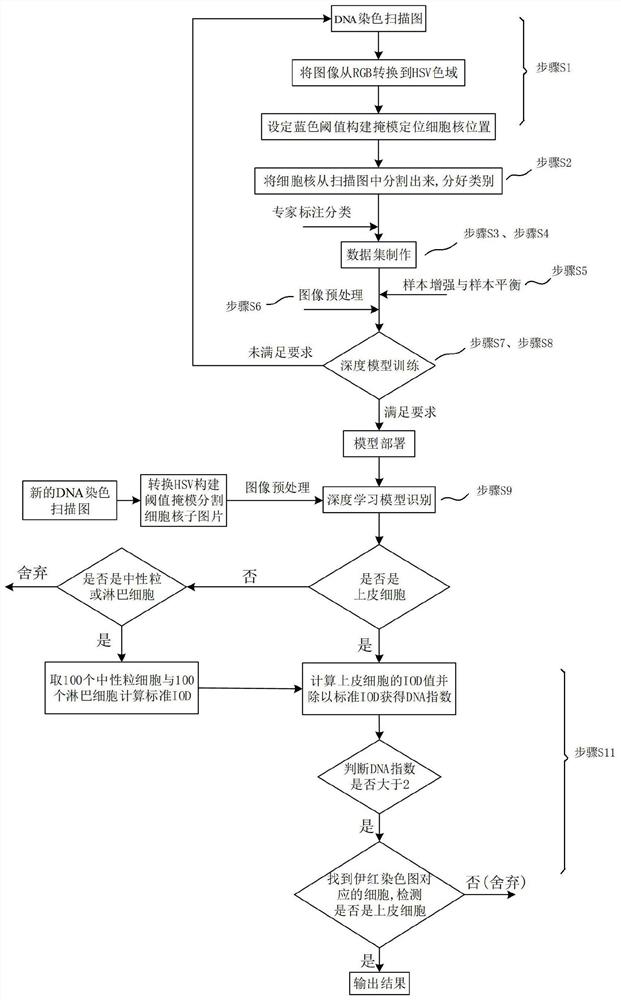
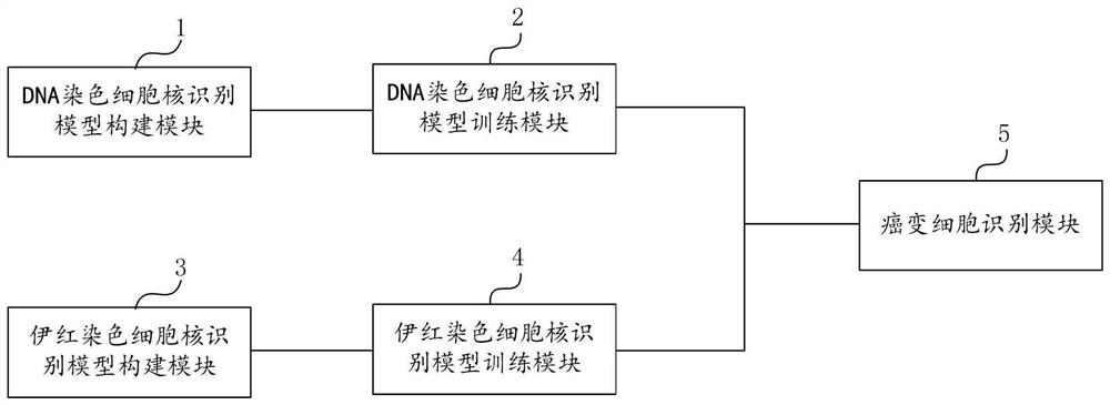
The invention discloses a cancerous cell detection method and system. The detection method comprises the following steps: constructing a model; constructing a DNA staining cell nucleus recognition model and an eosin staining cell nucleus recognition model; executing a cancerous cell recognition step; selecting suspected cancer cells by using the DNA staining cell nucleus recognition model, and correcting the suspected cancer cells by using the eosin staining cell nucleus recognition model. According to the cancerous cell detection method and system provided by the invention, the cancerous cell detection accuracy can be improved, and meanwhile, the cancerous cell detection efficiency can be improved.

Description

technical field [0001] The invention belongs to the technical field of cell detection and relates to a cell detection method, in particular to a cancerous cell detection method and system. Background technique [0002] At present, the traditional diagnosis is to make glass slides by staining, and the laboratory experts will view them one by one through a microscope. Due to the relatively large field of view of the slides and the large number of nuclei, it takes a long time for experts to determine the diagnosis of a pathological slide. , and there is a high possibility of missing possible cancer cells due to visual fatigue. [0003] In addition, because each examiner has his own subjective judgment, it is easy to cause the same film to be examined by different doctors, and there will be different results. [0004] In view of this, there is an urgent need to design a new cell detection method to overcome at least some of the above-mentioned defects in the existing cell detec...

Claims


Application Information

Patent Timeline
Patent Type & Authority Applications(China)
IPC IPC(8): G06T7/00G06T7/11G06T7/90G06K9/62
CPCG06T7/0012G06T7/11G06T7/90G06T2207/30024G06T2207/30096G06T2207/20081G06T2207/20084G06T2207/20132G06T2207/10056G06F18/24G06F18/214
Inventor 梅园
Owner 上海申挚医疗科技有限公司
Learn More