Check patentability & draft patents in minutes with Patsnap Eureka AI!

Medical ultrasonic teaching and training system

A training system, ultrasound technology, applied in the field of teaching and training system for simulating ultrasound images, can solve problems such as inability to effectively complete ultrasound diagnosis and treatment of patients, unfamiliar techniques, and poor results

Pending Publication Date: 2022-01-18
DONGGUAN TUNGWAH HOSPITAL +1
View PDF0 Cites 0 Cited by
  • Summary
  • Abstract
  • Description
  • Claims
  • Application Information

AI Technical Summary

Problems solved by technology

However, the operation of ultrasound image acquisition is a major difficulty in getting started with ultrasound diagnosis and treatment.
Ultrasonic slices are flexible and changeable, not limited to the three conventional slices of transverse, sagittal, and coronal slices. Therefore, for beginners, it is difficult to quickly understand the correspondence between the orientation of the ultrasound probe, anatomical slices, and images. ;For medical students or doctors with little clinical experience, it is difficult to have a personal and intuitive experience before taking up the job. The professional degree is not high during operation, and the technique is unfamiliar, and they cannot effectively complete the accurate ultrasound diagnosis and treatment of patients. The main solution at present is to spend A lot of time to practice under the guidance of the teacher; this kind of practice under the guidance of the teacher, the practice process is highly dependent on the teacher, and usually requires the hand-in-hand guidance of the teacher to complete and effectively practice in order to obtain better results ; Practitioners in the absence of guidance, independent practice takes a long time and the effect is poor; and there are big differences in the teaching quality of teachers with different qualifications

Method used


Image

  • Medical ultrasonic teaching and training system
  • Medical ultrasonic teaching and training system

Examples


Embodiment Construction

[0018] Embodiments of the present invention will now be described with reference to the drawings, in which like reference numerals represent like elements.

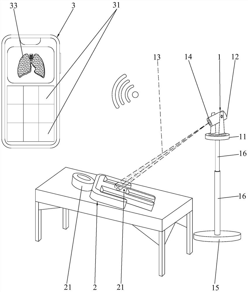
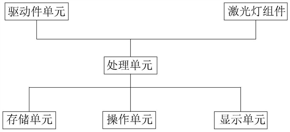
[0019] Such as figure 1 and figure 2As shown, the medical ultrasound teaching and training system of the present invention includes an ultrasound simulation device 1, a human body model 2 and a remote operation terminal 3; the ultrasound simulation device 1 includes a base 11 and a scanning part 12 and A driving part (not shown in the figure), the driving part drives the scanning part 12 to rotate and flip relative to the base 11 . Specifically, the driving part adopts an existing motor to drive the gear assembly, that is, the motor and the intermeshing gear assembly connected to the output shaft are arranged on the base 11 , and the output end of the gear assembly is connected to the scanning part 12 . The scanning unit 12 of the present invention includes a camera for positioning and a laser lamp assembly that emits ...


PUM


Abstract

The invention discloses a medical ultrasonic teaching and training system which comprises an ultrasonic simulation device, a human body model and a remote operation terminal, during use, an organ model needing to be trained is selected, an operation part corresponding to the organ model selected to be practiced is operated, and a processing unit controls a driving part to rotate or / and turn over to enable a camera to capture an identification part and stop. After the identification part is captured, the processing unit calls a motion path which is pre-stored in a storage unit and corresponds to the organ model selected to be practiced, and the processing unit controls the driving part to drive a scanning part to move to the position where a fan-shaped laser beam can scan the organ model selected to be practiced according to the called motion path; and a laser lamp assembly emits the fan-shaped laser beam to scan the organ model selected to be practiced, and a standard section image is displayed on a display unit at the same time. According to the invention, learning, observation and simulation are carried out in a whole-course self-service manner, so that the effect of ensuring the homogenization of teaching quality while a practicer completely learns autonomously is realized, and the practicer can quickly and effectively enter.

Description

technical field [0001] The invention relates to a medical teaching tool, in particular to a teaching and training system for simulating ultrasonic images. Background technique [0002] Ultrasonic diagnosis (ultrasonic diagnosis) is a method of diagnosis and treatment that applies ultrasonic detection technology to the human body, understands the data and shape of physiological or tissue structures, discovers diseases, and makes prompts. Ultrasound diagnosis is a non-invasive, painless, convenient, intuitive and effective means of examination. However, the operation of ultrasound image acquisition is a major difficulty in getting started with ultrasound diagnosis and treatment. Ultrasonic slices are flexible and changeable, not limited to the three conventional slices of transverse, sagittal, and coronal slices. Therefore, for beginners, it is difficult to quickly understand the correspondence between the orientation of the ultrasound probe, anatomical slices, and images. ;...

Claims


Application Information

Patent Timeline
Patent Type & Authority Applications(China)
IPC IPC(8): G09B9/00G09B5/02
CPCG09B9/00G09B5/02
Inventor 丁苇徐宝华黄小平叶茎宇
Owner DONGGUAN TUNGWAH HOSPITAL
Features
  • R&D
  • Intellectual Property
  • Life Sciences
  • Materials
  • Tech Scout
  • Unparalleled Data Quality
  • Higher Quality Content
  • 60% Fewer Hallucinations
Learn More