Imaging device applied to endoscope

An imaging device and endoscope technology, applied in the field of endoscope imaging, can solve the problems of being difficult to be observed, reduce the accuracy of endoscope imaging, and achieve the effect of accurate imaging results

Active Publication Date: 2022-02-15
WEST CHINA HOSPITAL SICHUAN UNIV
View PDF8 Cites 0 Cited by
  • Summary
  • Abstract
  • Description
  • Claims
  • Application Information

AI Technical Summary

Problems solved by technology

Then doctors observe the images obtained by the endoscope, and judge and identify whether the tissue is abnormal based on personal experience, but some tissue lesions may be difficult to observe, which greatly reduces the imaging accuracy of the endoscope

Method used


Image

  • Imaging device applied to endoscope
  • Imaging device applied to endoscope
  • Imaging device applied to endoscope

Examples


Embodiment Construction

[0036] In order to enable those skilled in the art to better understand the technical solutions in the present invention, the technical solutions in the embodiments of the present invention will be clearly and completely described below in conjunction with the drawings in the embodiments of the present invention. Obviously, the described The embodiments are only some of the embodiments of the present invention, not all of them. Based on the embodiments of the present invention, all other embodiments obtained by persons of ordinary skill in the art without making creative efforts shall fall within the protection scope of the present invention.

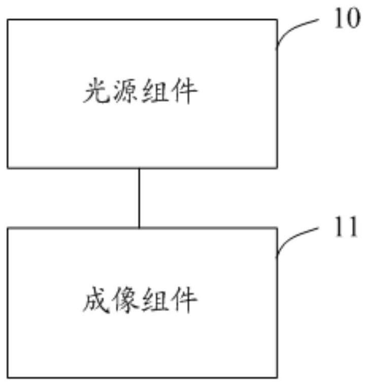
[0037] Please refer to figure 1 , figure 1 A schematic diagram of an imaging device applied to an endoscope provided in this embodiment, as shown in the figure, the imaging device applied to an endoscope includes a light source assembly 10 and an imaging assembly 11;

[0038] The light source assembly 10 is used to emit light of the f...


PUM


Abstract

The invention relates to an imaging device applied to an endoscope, a light source assembly emits first-wave-band light to a measured object, the light intensity is larger than zero when the first-wave-band light penetrates into a blood vessel in the measured object, and therefore the first-wave-band light can penetrate into the blood vessel in the measured object; due to the fact that the absorption capacity of the blood vessel to the first-wave-band light is different from the absorption capacity of other tissues in the measured object to the first-wave-band light, the vein blood vessel of the measured object can be shown in the image obtained by imaging the first-wave-band light reflected by the measured object through an imaging assembly. Therefore, the image representing the deep vein morphology and distribution of the detected object can be obtained, and the imaging result of optical imaging of the detected object is more accurate.

Description

technical field [0001] The invention relates to the field of endoscope imaging, in particular to an imaging device applied to endoscopes. Background technique [0002] Medical endoscope is a detection instrument that integrates optics, precision instruments, electronic circuits, and software. Detection of diseases, colon and other human internal organs. [0003] In the imaging process of the traditional endoscope, the white light source is irradiated to the surface of the human visceral tissue through the optical fiber channel of the endoscope, and the surface image of the human visceral tissue is obtained by using the imaging device of the endoscope. Furthermore, doctors observe the images obtained by the endoscope, and judge and identify whether the tissue is abnormal based on personal experience, but some tissue lesions may be difficult to observe, which greatly reduces the imaging accuracy of the endoscope. Contents of the invention [0004] The object of the present...

Claims


Application Information

Patent Timeline
Patent Type & Authority Applications(China)
IPC IPC(8): A61B1/04A61B1/06A61B5/00
CPCA61B1/04A61B1/06A61B5/0084A61B5/0075A61B5/0033A61B5/489
Inventor 胡兵陈柳香
Owner WEST CHINA HOSPITAL SICHUAN UNIV
Why Eureka
Try Eureka