Nasopharyngeal carcinoma identification and tumor segmentation method and system based on MR image

A nasopharyngeal carcinoma and image technology, applied in the field of nasopharyngeal carcinoma identification and tumor segmentation method and system, can solve the problems of optimization, single function, and inability to apply at the same time, so as to consolidate robustness, enhance generalization ability, and improve segmentation effect of effect

Pending Publication Date: 2022-03-08
SUN YAT SEN UNIV CANCER CENT
View PDF0 Cites 0 Cited by
  • Summary
  • Abstract
  • Description
  • Claims
  • Application Information

AI Technical Summary

Problems solved by technology

[0004] The present invention provides a nasopharyngeal carcinoma identification and tumor segmentation method and system based on MR images to solve the problem that the existing nasopharyngeal carcinoma identification and tumor segmentation methods and systems have single functions and can only perform tumor segmentation on nasopharyngeal carcinoma MR images However, the 3D deep learning model for nasopharyngeal carcinoma identification and tumor segmentation has not been optimized to further improve the performance of the model, and it cannot be applied to the technical problems of MR plain scan sequence images and MR enhanced sequence images at the same time

Method used


Image

  • Nasopharyngeal carcinoma identification and tumor segmentation method and system based on MR image
  • Nasopharyngeal carcinoma identification and tumor segmentation method and system based on MR image
  • Nasopharyngeal carcinoma identification and tumor segmentation method and system based on MR image

Examples


Embodiment 1

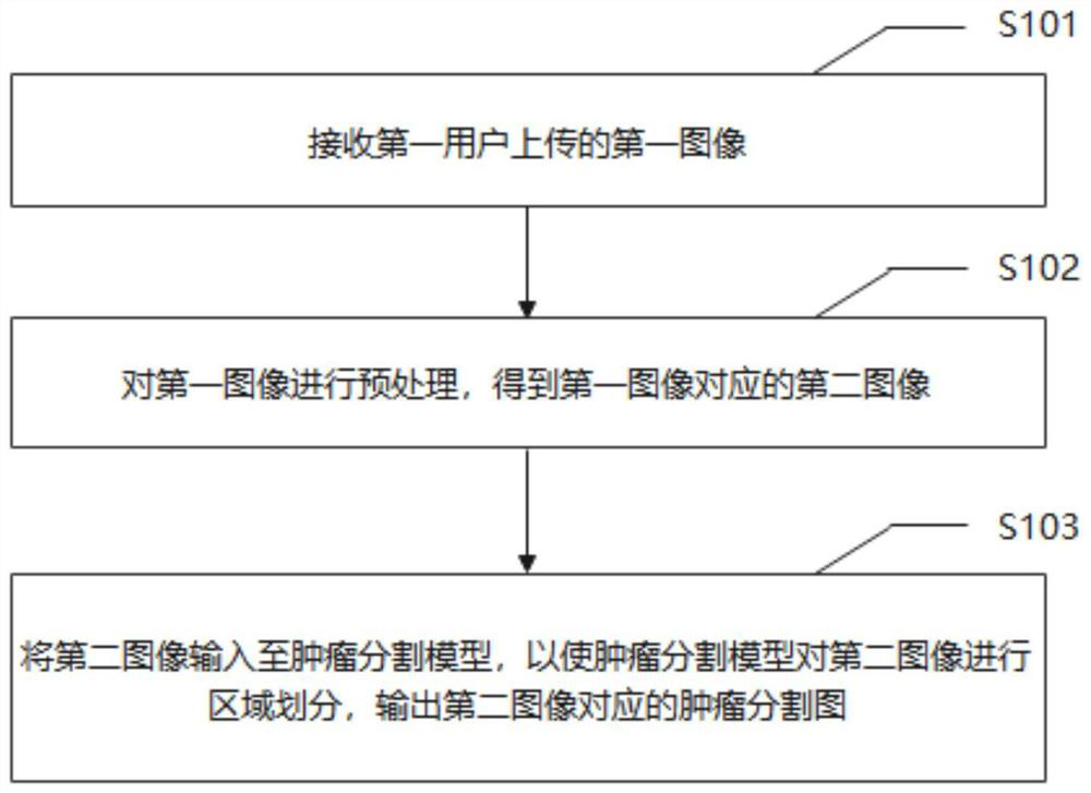
[0055] Please refer to figure 1 , an MR image-based nasopharyngeal carcinoma identification and tumor segmentation method provided in an embodiment of the present invention, comprising:

[0056] S101: Receive a first image uploaded by a first user; wherein, the first image includes an MR unenhanced sequence image or an MR enhanced sequence image, and the first user may be a clinician or a patient.

[0057] S102: Perform preprocessing on the first image to obtain a second image corresponding to the first image.

[0058] In this embodiment, further, the preprocessing of the first image to obtain the second image of the first image is specifically:

[0059] The first image is preprocessed by image data cleaning, data normalization, plain scan and enhanced image registration, to obtain a second image corresponding to the first image.

[0060] It should be noted that, by performing preprocessing on the first image, the first image is converted into a second image corresponding to...


PUM


Abstract

The invention discloses a nasopharyngeal carcinoma recognition and tumor segmentation method and system based on an MR image. The method comprises the following steps: receiving a first image uploaded by a first user; wherein the first image comprises an MR plain-scan sequence image or an MR enhanced sequence image; preprocessing the first image to obtain a second image corresponding to the first image; inputting the second image into a tumor segmentation model to enable the tumor segmentation model to carry out region division on the second image, and outputting a tumor segmentation map corresponding to the second image; wherein the tumor segmentation model is obtained by training a three-dimensional convolutional neural network model according to a plurality of first nasopharyngeal benign and malignant MR image data and a plurality of corresponding second nasopharyngeal benign and malignant MR image data with tumor markers. According to the method, the tumor segmentation model suitable for the MR plain scanning sequence and the MR enhanced sequence is adopted, nasopharyngeal carcinoma recognition and tumor segmentation of the MR image are achieved, the model is optimized through data obtained through segmentation mistakes and omissions, and the segmentation effect of the image is improved.

Description

technical field [0001] The invention relates to the field of medical image processing, in particular to a method and system for nasopharyngeal carcinoma identification and tumor segmentation based on MR images. Background technique [0002] Nasopharyngeal carcinoma is a malignant tumor originating from the epithelial cells of the nasopharynx, which has great potential for invasion and metastasis. Because the anatomical location of nasopharyngeal carcinoma is not obvious and the symptoms are nonspecific, most patients are diagnosed with advanced disease. On the other hand, the complex skull base anatomy increases the difficulty of NPC segmentation, thereby reducing the accuracy of disease staging and compliance with radiotherapy. Magnetic Resonance imaging (MRI) is the preferred imaging modality for identification, staging, curative effect evaluation and post-treatment follow-up of nasopharyngeal carcinoma patients due to its resolution of soft tissue, detection sensitivity ...

Claims


Application Information

Patent Timeline
IPC IPC(8): G06T7/00G06T7/11G06N3/04G06N3/08
CPCG06T7/0012G06T7/11G06N3/08G06T2207/20081G06T2207/20084G06T2207/30096G06T2207/10088G06N3/045
Inventor 邓一术李超峰经秉中李彬陈浩华
Owner SUN YAT SEN UNIV CANCER CENT
Why Eureka
Try Eureka