Tumor risk auxiliary diagnosis equipment based on radiomics

A radiomics, auxiliary diagnosis technology, applied in the field of medical imaging, can solve the problems of limited space, affecting doctors' observation, inconvenient placement and movement of photo lights, etc., to achieve the effect of portability

Inactive Publication Date: 2022-04-29
山东承势电子科技有限公司
View PDF0 Cites 0 Cited by
  • Summary
  • Abstract
  • Description
  • Claims
  • Application Information

AI Technical Summary

Problems solved by technology

[0003] At present, most of the photo lights are fixed or desktop, which are prone to collisions during the moving process and are inconvenient to move. Some smaller photo lights are easy to carry and move, but the photo area is small, which affects the doctor's observation. The area, the venue is limited, it is not convenient to place and move the photo light

Method used


Image

  • Tumor risk auxiliary diagnosis equipment based on radiomics
  • Tumor risk auxiliary diagnosis equipment based on radiomics
  • Tumor risk auxiliary diagnosis equipment based on radiomics

Examples


Embodiment Construction

[0028] In order to make the technical means, creative features, goals and effects achieved by the present invention easy to understand, the present invention will be further described below in conjunction with specific embodiments.

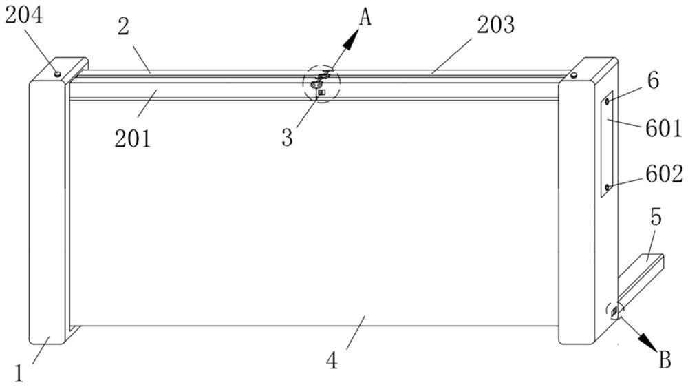
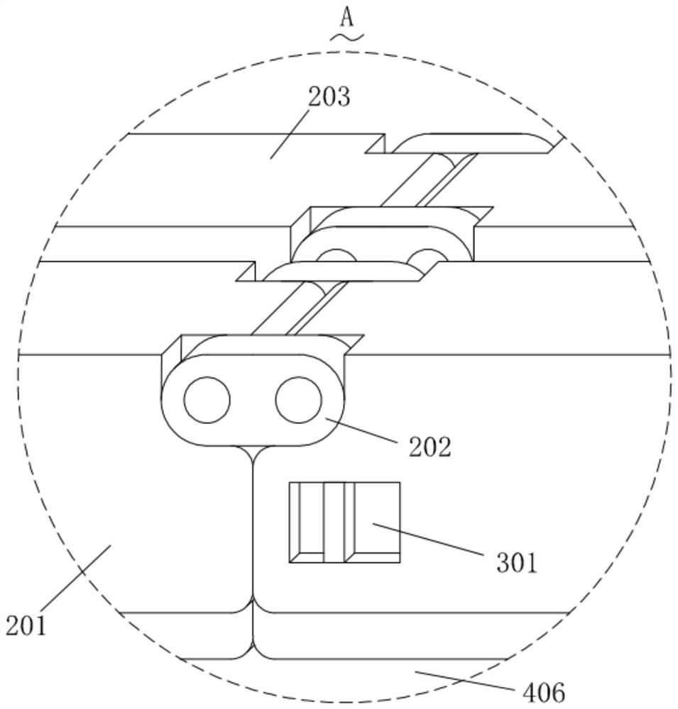
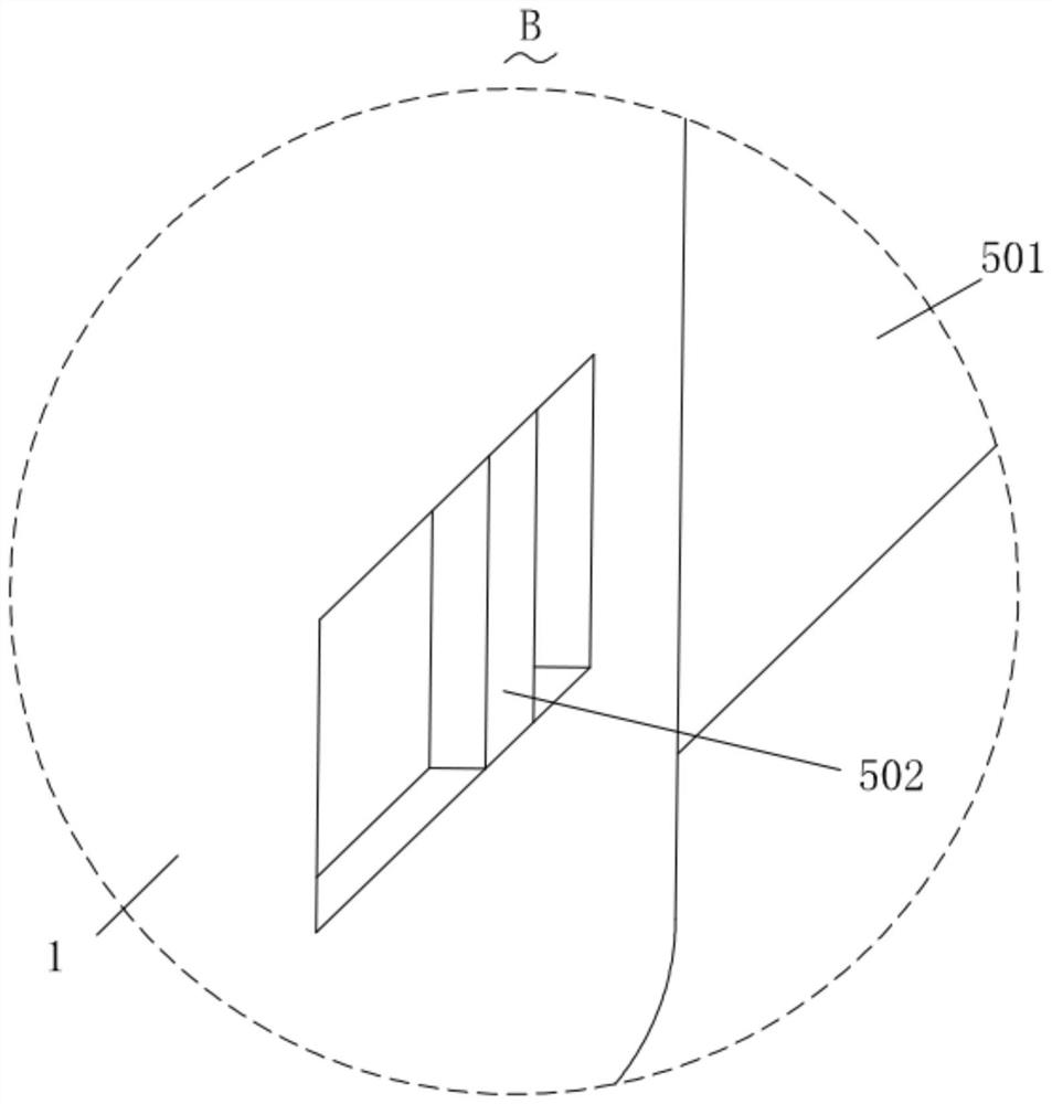
[0029] Such as Figure 1-Figure 10 As shown, the radiomics-based tumor risk auxiliary diagnosis device of the present invention includes a pair of fixed plates 1, and a pair of fixed plates 1 are connected with an unfolding structure 2 for expanding and supporting the fixed plate 1. The expansion structure 2 is connected with a position-limiting structure 3 for limiting the expansion structure 2, and a pair of the fixing plates 1 are connected with a lighting structure 4 for providing lighting, and the fixing plate 1 is connected with a lighting structure 4 for providing lighting. The structure 4 provides a power supply structure 6 for power supply, the lighting structure 4 is connected with a fixing structure 7 for fixing images, and the fixing p...


PUM


Abstract

The invention relates to the technical field of medical imaging, in particular to imaging omics-based tumor risk auxiliary diagnosis equipment which comprises a pair of fixing plates, an unfolding structure is connected to the pair of fixing plates, a limiting structure is connected to the unfolding structure, a lighting structure is connected to the pair of fixing plates, and the limiting structure is connected to the lighting structure. The fixing plate is connected with a power supply structure used for providing power for the lighting structure, the lighting structure is connected with a fixing structure used for fixing an image film, and the fixing plate is connected with a supporting structure used for supporting the fixing plate. The device is convenient to unfold through the unfolding structure and convenient to carry when folded, under the action of the limiting structure, the unfolding structure can be limited, the image film can be conveniently fixed through the fixing structure, the image film can be illuminated through the illumination structure, the power supply structure can provide power for the illumination structure, and the supporting structure can support the device. The device toppling is avoided.

Description

technical field [0001] The invention relates to the technical field of medical imaging, in particular to an auxiliary diagnosis device for tumor risk based on radiomics. Background technique [0002] In the process of tumor diagnosis, CT and other equipment are usually used to scan and image the patient's part. When analyzing the image, it is necessary to take a photo through a photo lamp, so as to facilitate the observation of the image. [0003] At present, most of the photo lights are fixed or desktop, which are prone to collisions during the moving process and are inconvenient to move. Some smaller photo lights are easy to carry and move, but the photo area is small, which affects the doctor's observation. The area and venue are limited, which is not convenient for the placement and movement of photo lights. Contents of the invention [0004] Aiming at the problems in the prior art, the present invention provides an auxiliary diagnosis device for tumor risk based on r...

Claims


Application Information

Patent Timeline
Patent Type & Authority Applications(China)
IPC IPC(8): G02B27/04G02B27/02
CPCG02B27/04G02B27/028G02B27/024
Inventor 付树军齐泽荣胡明征
Owner 山东承势电子科技有限公司
Why Eureka
Try Eureka