Endometrial cancer cell detection method, system and equipment and storage medium

A technology for endometrium and detection methods, applied in the field of systems, equipment and storage media, and endometrial cancer cell detection methods, can solve the problems of slow time-consuming, low precision, and strong subjectivity, and reduce labor loss and price Low cost and effect of reducing economic burden

Pending Publication Date: 2022-06-03
XI AN JIAOTONG UNIV +1
View PDF0 Cites 1 Cited by
  • Summary
  • Abstract
  • Description
  • Claims
  • Application Information

AI Technical Summary

Problems solved by technology

[0006] The purpose of the present invention is to solve the problems of slow time-consuming, low precision and strong subjectivity in the pathological image diagnosis process of endometrial cancer cells, and provide a method, system, equipment and storage medium for detecting endometrial cancer cells

Method used


Image

  • Endometrial cancer cell detection method, system and equipment and storage medium
  • Endometrial cancer cell detection method, system and equipment and storage medium
  • Endometrial cancer cell detection method, system and equipment and storage medium

Examples


Embodiment

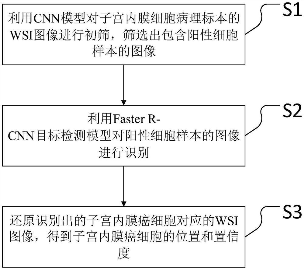
[0052] The present invention is based on the Faster R-CNN intelligent detection method for endometrial cancer cells, and the specific identification steps are as follows:

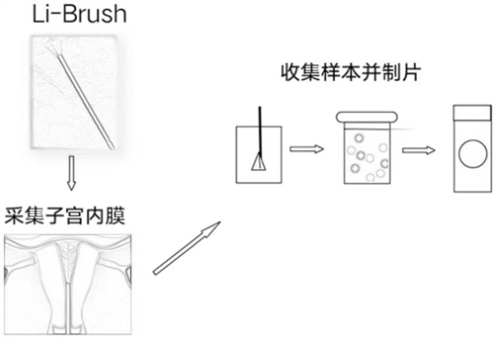
[0053] Step 1: Collect pathological specimens of endometrial cells from patients to be examined clinically through the Li-brush endometrial cell collector, and scan the specimens with a unique scanner to produce WSI containing hundreds of millions of pixels.

[0054] First, the user brushes the patient's endometrial cell biological sample with Li-brush, and then places the prepared glass slide with the cell sample on the stage for scanning, and the patient's endometrial can be obtained. Full picture of cytopathology.

[0055] This step is the basis for the network training of the subsequent steps. A sample with high definition and uniform staining is of great significance for computer recognition. Therefore, we have formulated a standardized and unified collection and production process:

[0056] Sample co...


PUM


Abstract

The invention discloses an endometrial cancer cell detection method, system and equipment and a storage medium. The method comprises the following steps: preparing an endometrial cell pathological section; preliminarily screening images containing positive cells by using CNN; performing intelligent detection on cancer cells by using the improved Faster R-CNN target detection model; and performing statistics on each index, sending the WSI generated blocks into network prediction, and then merging identification results. In the target acquisition part, a data set is normalized and expanded by using a self-adaptive random clipping method on the aspect of data; in the model, an improved CNN model and a two-stage Faster RCNN model are taken as basic models, diagnosis experience of doctors is introduced, and diagnosis indexes are extracted to jointly act to train a more accurate model. The accuracy rate of the endometrial cancer cell detection method is higher than that of a classification method, meanwhile, the confidence coefficient and the position information of the WSI can be given, and in addition, the invention further provides a preparation method of a more effective endometrial cell pathological section image data set. The method is applied to the field of medical image target detection.

Description

technical field [0001] The invention belongs to the technical field of AI medical imaging, and relates to an endometrial cancer cell detection method, system, equipment and storage medium. Background technique [0002] Endometrial cancer is a group of epithelial malignant tumors that occur in the endometrium. It is one of the three major malignant tumors of the female reproductive system. There are more than 378,000 patients in the world every year, and its incidence is increasing worldwide. and tend to be younger. In addition, studies have shown that the 5-year survival rate for early-stage endometrial cancer with appropriate treatment is over 85%. Early screening and early diagnosis of precancerous lesions of endometrial cancer can greatly reduce its incidence and mortality. [0003] Computer-aided diagnosis systems can help doctors effectively analyze and evaluate a large number of medical images. It has been applied to the radiodiagnosis of some common cancers such as ...

Claims


Application Information

Patent Timeline
IPC IPC(8): G06K9/62G06N3/04G06N3/08G06T7/00G06T7/73
CPCG06T7/0012G06T7/73G06N3/08G06T2207/10061G06T2207/20081G06T2207/20084G06T2207/30096G06N3/045G06F18/23213G06F18/24
Inventor 钟德星李奇灵谢中林赵惊涛
Owner XI AN JIAOTONG UNIV
Why Eureka
Try Eureka