
Breast biopsy system and methods

a biopsy system and a technology for removing blood, applied in the field of methods and devices, can solve the problems of inability to acquire tissue samples having a cross-section larger than, no single procedure is ideal for all cases, etc., and achieve the effect of minimizing the risk of migration and high likelihood of “clean” margins

Inactive Publication Date: 2005-01-13
SENORX
View PDF83 Cites 134 Cited by
  • Summary
  • Abstract
  • Description
  • Claims
  • Application Information

AI Technical Summary

Benefits of technology

The present invention provides an apparatus and method for accurately isolating a target lesion, resulting in a high likelihood of "clean" margins. This advantageously allows for both diagnosis and treatment of a malignant lesion with only a single percutaneous procedure, with no follow-up percutaneous or surgical procedure required, while minimizing the risk of migration of possibly cancerous cells from the lesion to surrounding tissue or the bloodstream. The invention includes a biopsy instrument with a cutting element that can be actuated between a radially retracted position and a radially extended position, and an encapsulating element that can be actuated between a radially retracted position and a radially extended position. The instrument is inserted into the patient's body and the cutting element is expanded and rotated to create a peripheral boundary about the tissue specimen, allowing for precise isolation from surrounding tissue. The invention provides a minimally invasive and efficient approach to diagnostic and therapeutic procedures.

Problems solved by technology

The type of biopsy utilized depends in large part on circumstances present with respect to the patient, including the location of the lesion(s) within the body, and no single procedure is ideal for all cases.
A disadvantage of this type of device, however, is the inability to acquire a tissue sample having a cross-section larger than that of the cannula through which the sample will be removed.
Additionally, it is difficult, using such a device, which obtains cylindrical shaped specimens, to determine whether an entire lesion of interest is being removed or whether a further procedure will be necessary.

Method used


Image

  • Breast biopsy system and methods
  • Breast biopsy system and methods
  • Breast biopsy system and methods

Examples


Embodiment Construction

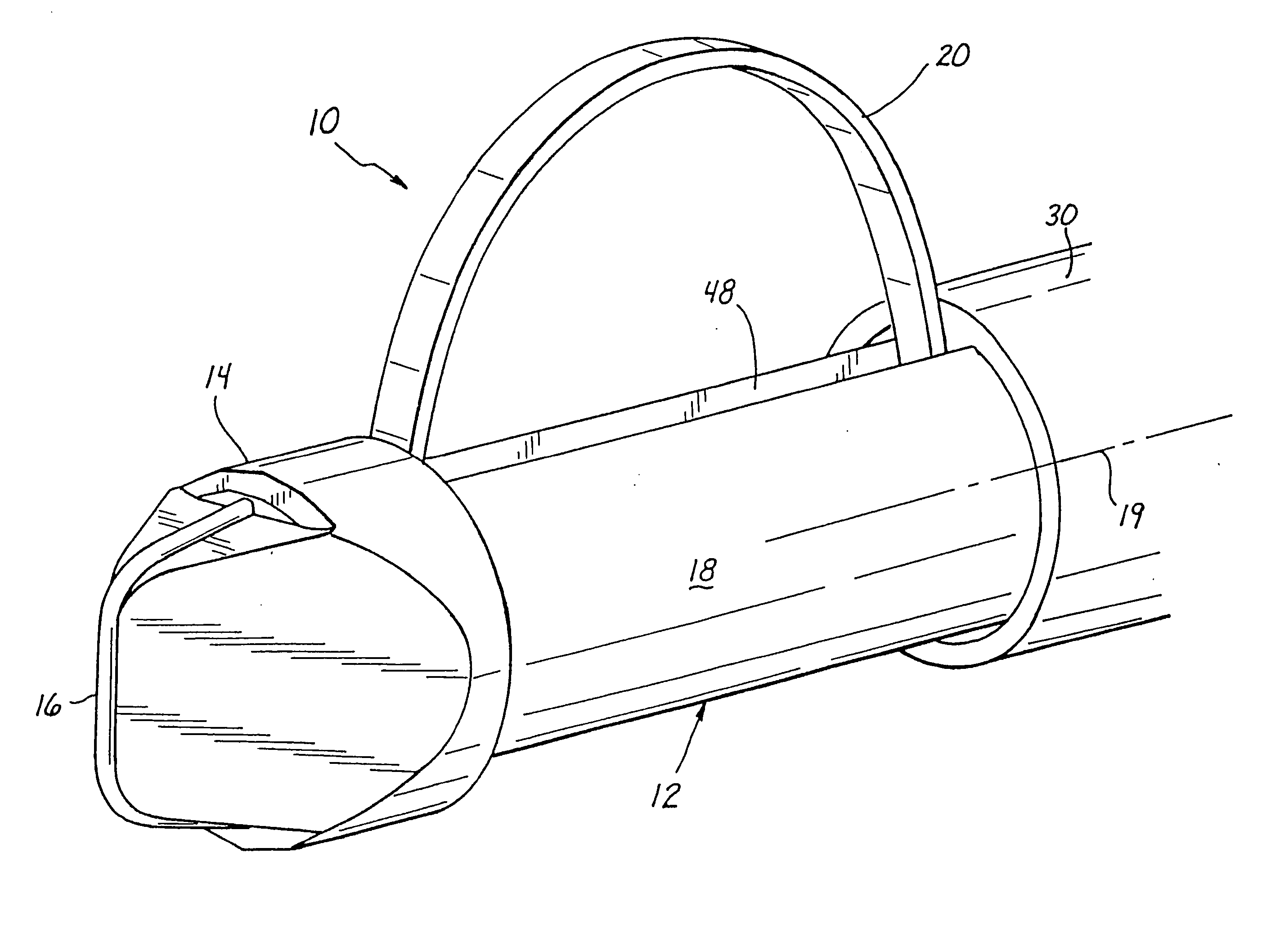
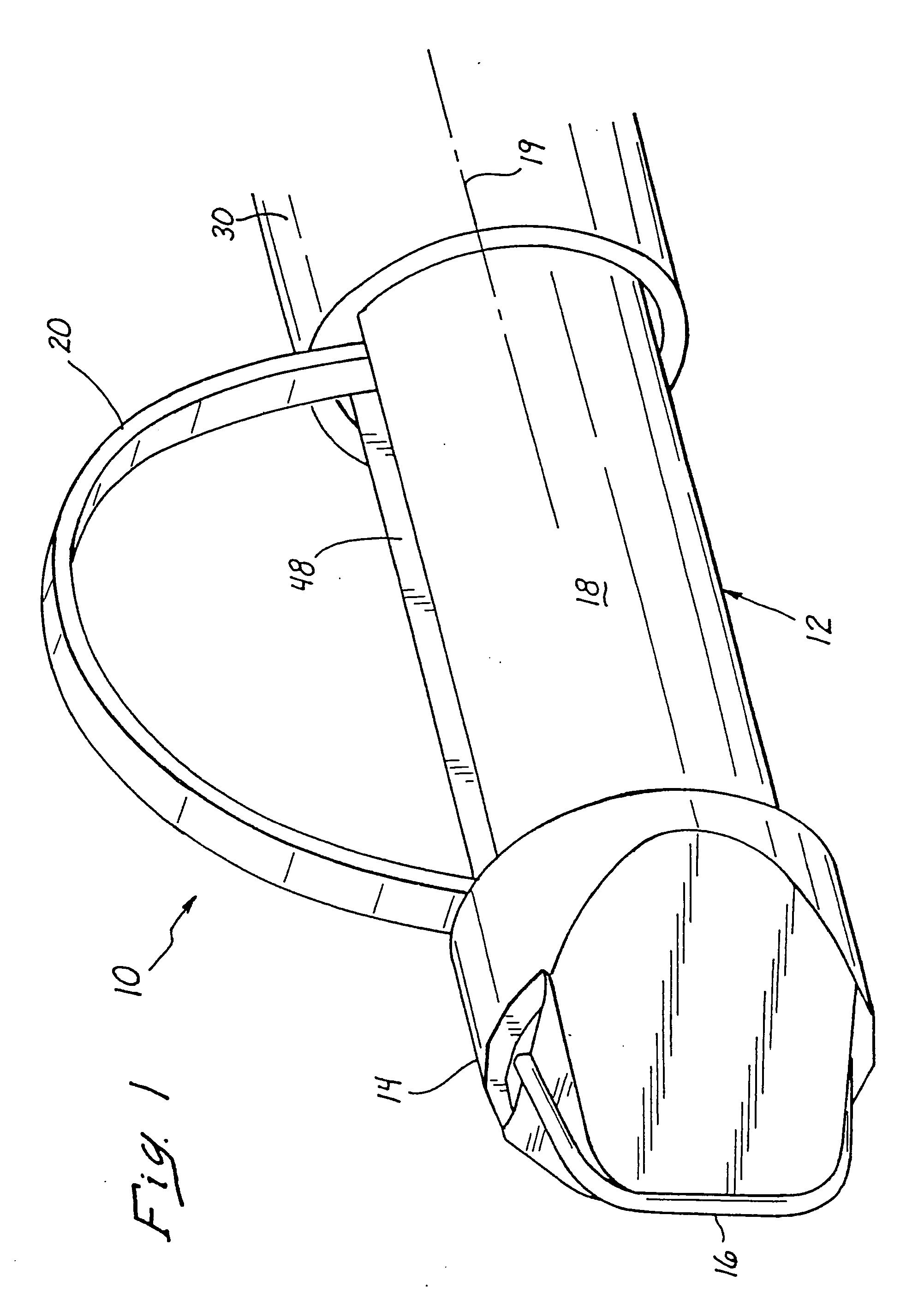
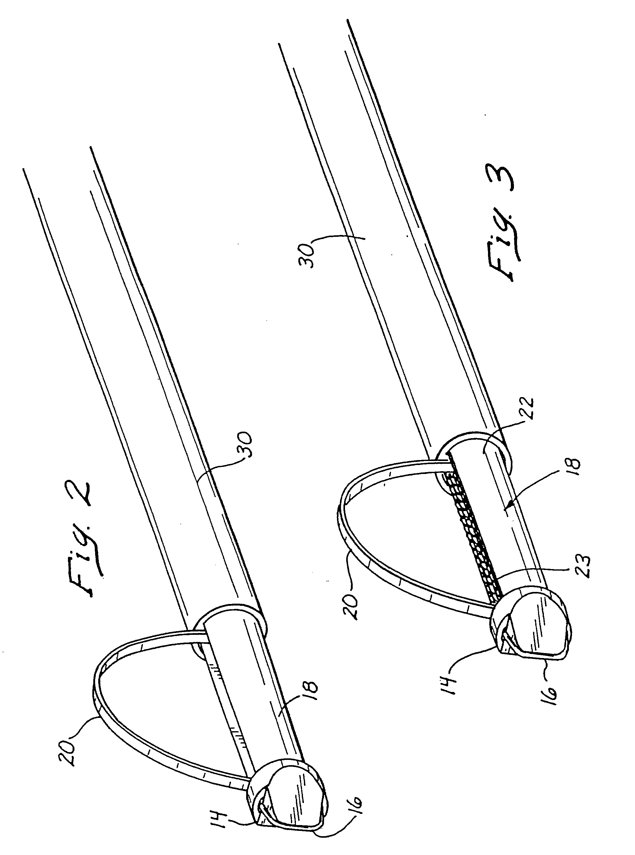
[0032] Referring now more particularly to FIG. 1, there is shown the distal end 12 of a first preferred embodiment of an inventive tissue retrieval or biopsy instrument 10. The distal end 12 preferably comprises a disposable wand portion, including a distal tip 14. The tip 14 may comprise a conventional trocar tip, or, preferably, may include an electrosurgical (RF) element or wire 16 which may be energized by a conventional electrosurgical generator (not shown) in order to facilitate tissue cutting and consequent advancement of the instrument 10 to a predetermined tissue site in the patient's body.

[0033] Proximally of the tip 14 is a shaft 18, preferably lying along an axis 19 (FIG. 1) of the instrument, on which is disposed a cutting element or wire 20. This wire 20 is disposed axially along the length of the shaft 18 in its retracted position (not shown), but may be deployed radially outwardly, as shown in FIG. 1. The element 20 is preferably comprised of a wire or rectangular b...


PUM


Abstract

An apparatus and method are provided for precisely isolating a target lesion in a patient's body tissue, resulting in a high likelihood of “clean” margins about the lesion when it is removed for diagnosis and / or therapy. This approach advantageously will often result in the ability to both diagnose and treat a malignant lesion with only a single percutaneous procedure, with no follow-up percutaneous or surgical procedure required, while minimizing the risk of migration of possibly cancerous cells from the lesion to surrounding tissue or the bloodstream. In particular, the apparatus comprises a biopsy instrument having a distal end adapted for entry into the patient's body, a longitudinal shaft, and a cutting element disposed along the shaft. The cutting element is actuatable between a radially retracted position and a radially extended position. Advantageously, the instrument is rotatable about its axis in the radially extended position to isolate a desired tissue specimen from surrounding tissue by defining a peripheral margin about the tissue specimen. Once the tissue specimen is isolated, it may be segmented by further manipulation of the cutting element, after which the tissue segments are preferably individually removed from the patient's body through a cannula or the like. Alternatively, the specimen may be encapsulated and removed as an intact piece.

Description

FIELD OF THE INVENTION [0001] The present invention relates to methods and devices for removing tissue samples, and more specifically to improved instruments and methods for acquiring soft body tissue. BACKGROUND OF THE INVENTION [0002] It is often desirable and frequently necessary to sample or remove a portion of tissue from humans and other animals, particularly in the diagnosis and treatment of patients with cancerous tumors, pre-malignant conditions, and other diseases or disorders. [0003] Typically, in the case of cancer, particularly cancer of the breast, there is a great emphasis on early detection and diagnosis through the use of screening modalities, such as physical examination, and particularly mammography, which is capable of detecting very small abnormalities, often nonpalpable. When the physician establishes by means of a mammogram or other screening modality, such as ultrasound, that suspicious circumstances exist, a biopsy must be performed to capture tissue for a d...

Claims


Application Information

Patent Timeline
Patent Type & Authority Applications(United States)
IPC IPC(8): A61B10/00A61B10/02A61B17/00A61B17/22A61B17/32A61B17/34A61B18/12A61B18/14A61B19/00
CPCA61B10/02A61B2019/5408A61B17/00491A61B17/32056A61B17/320725A61B17/3417A61B18/14A61B18/1487A61B18/1492A61B19/5225A61B2017/3488A61B2018/00208A61B2018/00214A61B2018/00267A61B2018/00333A61B2018/00898A61B2018/0091A61B2018/00916A61B2018/1253A61B2018/126A61B2018/1407A61B2018/1475A61B2018/162A61B2019/205A61B10/0266A61B90/17A61B90/37A61B2090/3908
Inventor BURBANK, FRED H.JONES, MICHAEL L.LUBOCK, PAUL
Owner SENORX
Learn More