
Ultrasonic imaging device

An ultrasonic imaging and ultrasonic echo technology, applied in the medical field, can solve the problems of consuming clinical examination time, time-consuming and laborious, lack of consistency in image display results, etc., to reduce clinical examination time, improve accuracy, and achieve good consistency. Effect

Pending Publication Date: 2022-06-21
GUANGDONG WOMEN & CHILDREN HOSPITAL +1
View PDF0 Cites 0 Cited by
  • Summary
  • Abstract
  • Description
  • Claims
  • Application Information

AI Technical Summary

Problems solved by technology

The whole process requires doctors to have rich clinical experience, and it is time-consuming and laborious
[0005] Therefore, in the prior art, when detecting the spine, the use of three-dimensional imaging relies too much on the experience and operation of doctors, consumes time for clinical examination, and the obtained image display results also lack good consistency.

Method used


Image

  • Ultrasonic imaging device
  • Ultrasonic imaging device
  • Ultrasonic imaging device

Examples


Embodiment Construction

[0023] In order to make the objects, technical solutions and advantages of the present invention more apparent, exemplary embodiments according to the present invention will be described in detail below with reference to the accompanying drawings. Obviously, the described embodiments are only some of the embodiments of the present invention, not all of the embodiments of the present invention, and it should be understood that the present invention is not limited by the example embodiments described herein. Based on the embodiments of the present invention described in the present invention, all other embodiments obtained by those skilled in the art without creative efforts shall fall within the protection scope of the present invention.

[0024] In a 3D imaging system, the 3D visualization information usually includes the display of sectional (or cross-section, MultiplePlanner Rendering, MPR) images and the display of stereoscopic images (Volume Rendering, VR). The image obtai...


PUM


Abstract

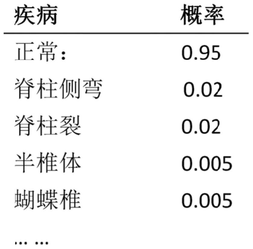
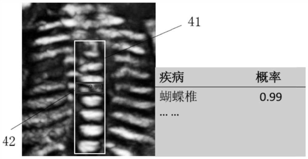
The invention provides an ultrasonic imaging device. The ultrasonic imaging device comprises a probe; the transmitting circuit is used for exciting the probe to transmit ultrasonic waves to the spine for body scanning; the receiving circuit is used for receiving the ultrasonic echo returned from the spine through the probe so as to obtain an ultrasonic echo signal; the processor is used for acquiring three-dimensional body data of the spine according to the ultrasonic echo signal; identifying a key anatomical structure of the spine from the three-dimensional body data of the spine to obtain key anatomical structure data of the spine; adjusting the three-dimensional body data to a preset angle according to the key anatomical structure to obtain standard three-dimensional body data, and obtaining a standard image of the spine based on the standard three-dimensional body data; and inputting at least one of the standard image and the spine key anatomical region data into a detection neural network to obtain a detection result of the spine, and a display for displaying the standard image of the spine and the detection result of the spine. According to the device, the detection result of the spine is visually displayed, the detection time is saved, and the accuracy of the detection result is improved.

Description

technical field [0001] The present invention relates to the field of medical technology, and more particularly to ultrasound imaging. Background technique [0002] Ultrasonography has been widely used in clinical examination due to its advantages of safety, convenience, non-radiation, and low cost, and has become one of the main auxiliary means for doctors to diagnose diseases. Ultrasound avoids the influence of X-rays on the human body, and its application value Significantly better than other imaging equipment. [0003] The spine is a very important structure in the human body, especially an important part of judging fetal development in prenatal screening. In recent years, 3D ultrasound has been widely used in human body examination. Its advantage is that 3D volume data of the region of interest can be obtained through one scan, and any section in the volume data can be displayed. The images are intuitive and can help doctors to locate more accurately Abnormal segment o...

Claims


Application Information

Patent Timeline
Patent Type & Authority Applications(China)
IPC IPC(8): A61B8/08G06V20/64G06V10/764G06K9/62
CPCA61B8/0866A61B8/0875A61B8/483A61B8/5215A61B8/5223G06F18/241
Inventor 尚宁郭爽萍贾洪飞梁天柱林穆清
Owner GUANGDONG WOMEN & CHILDREN HOSPITAL
Learn More