
Injection device for salpingography

A technology of injection device and fallopian tube, which can be applied to balloon catheters, medical devices, other medical devices, etc., and can solve problems such as troublesome operation.

Pending Publication Date: 2022-07-22
郑州大学第三附属医院
View PDF0 Cites 1 Cited by
  • Summary
  • Abstract
  • Description
  • Claims
  • Application Information

AI Technical Summary

Problems solved by technology

[0003] During the above-mentioned whole operation process of placing the catheter, the medical personnel need to hold the catheter in one hand and the tweezers in the other hand. When other operations need to be performed, the rest of the medical personnel are required to help, and the operation is more troublesome.

Method used


Image

  • Injection device for salpingography
  • Injection device for salpingography
  • Injection device for salpingography

Examples


Embodiment Construction

[0034] Attached to the following Figure 1-7 This application will be described in further detail.

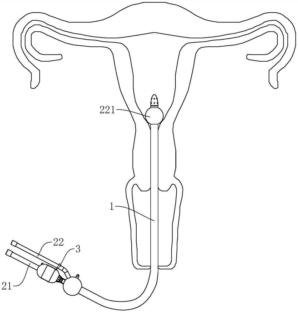
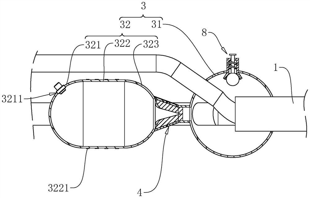
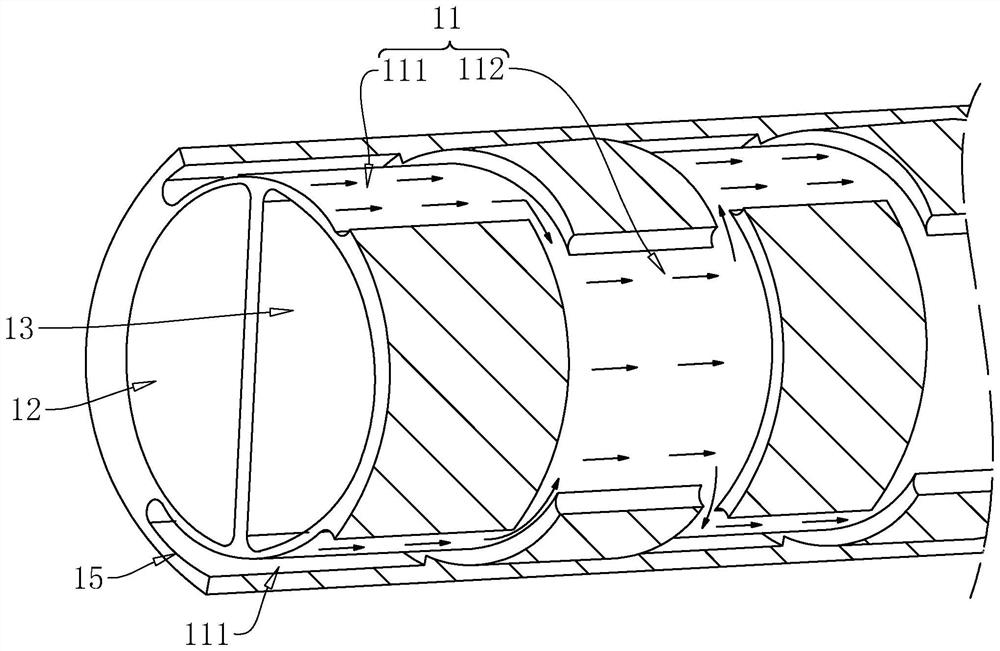
[0035] The embodiment of the present application discloses an injection device for salpingography. refer to Figure 1 to Figure 3 An injection device for salpingography includes a catheter body 1, a gas storage cavity 11 is arranged in the catheter body 1 along its own length direction, and gas is filled into the air storage cavity 11, and the catheter body 1 is in the air storage cavity 11. The strength increases under the action of the gas, and it is not easy to bend, so that the medical staff can directly insert the catheter into the uterine cavity of the patient, which is more convenient when performing the contrast agent injection operation.

[0036] refer to figure 1 and image 3In this embodiment, a two-lumen and one-balloon tube is used as an example. In other embodiments, the catheter body 1 can also be set on a three-lumen, two-balloon tube or other catheters that...


PUM


Abstract

The invention relates to the field of medical instruments, in particular to an injection device for oviduct radiography, which comprises a catheter body, a radiography channel is arranged in the catheter body, an insertion end and an injection end are arranged on the catheter body, a gas storage cavity channel for accommodating gas is arranged in the catheter body, and the gas storage cavity channel is communicated with the insertion end and the injection end. The air storage cavity extends to the insertion end from the injection end of the catheter body, and an inflation assembly used for inflating the air storage cavity is arranged at the end, close to the injection end, of the catheter body. In the application, the catheter body can be straightened and is not easy to bend by introducing gas into the gas storage channel, and medical staff can insert the catheter body into the uterine cavity of a patient more conveniently, so that the medical staff can perform contrast agent injection operation more conveniently.

Description

technical field [0001] The invention relates to the field of medical instruments, in particular to an injection device for salpingography. Background technique [0002] Salpingography is a method of injecting contrast agent into the uterine cavity and fallopian tube, using X-ray fluoroscopy and photography, according to the development of contrast agent in the fallopian tube and pelvis to understand whether the fallopian tube is unobstructed, the location of obstruction and the shape of the uterine cavity. When the contrast agent is injected, the patient lies supine on the examination bed, and the medical staff needs to insert the catheter into the uterine cavity through the patient's vaginal opening. The catheter is provided with a contrast agent channel and a balloon channel. The contrast agent channel is used for contrast agent injection. One end of the balloon channel is connected to A spherical balloon is provided; in the specific operation, the medical staff needs to f...

Claims


Application Information

Patent Timeline
IPC IPC(8): A61M31/00A61M25/10
CPCA61M31/00A61M25/1011A61M25/0026A61M2210/1433A61M2025/1013A61M2210/005
Inventor 张红彬栗河舟
Owner 郑州大学第三附属医院
Learn More