
SENet-based gastric cancer pathological section image segmentation prediction method

A technology of pathological sectioning and image segmentation, which is applied in the field of image processing, can solve the problems of high complexity and poor detection effect of small lesions, achieve the effect of increasing the depth of representation, improving the accuracy of detection, and solving the effects of computing resources

Pending Publication Date: 2022-08-05
SOUTH CHINA UNIV OF TECH
View PDF0 Cites 0 Cited by
  • Summary
  • Abstract
  • Description
  • Claims
  • Application Information

AI Technical Summary

Problems solved by technology

[0006] In order to overcome the defects and deficiencies in the prior art, the present invention provides a SENet-based method for segmenting and predicting gastric cancer pathological slice images, which solves the problems of high complexity and poor detection effect on tiny lesions in existing models, and can be used for gastric cancer Segmentation of full-field pathological slice images

Method used


Image

  • SENet-based gastric cancer pathological section image segmentation prediction method
  • SENet-based gastric cancer pathological section image segmentation prediction method
  • SENet-based gastric cancer pathological section image segmentation prediction method

Examples


Embodiment 1

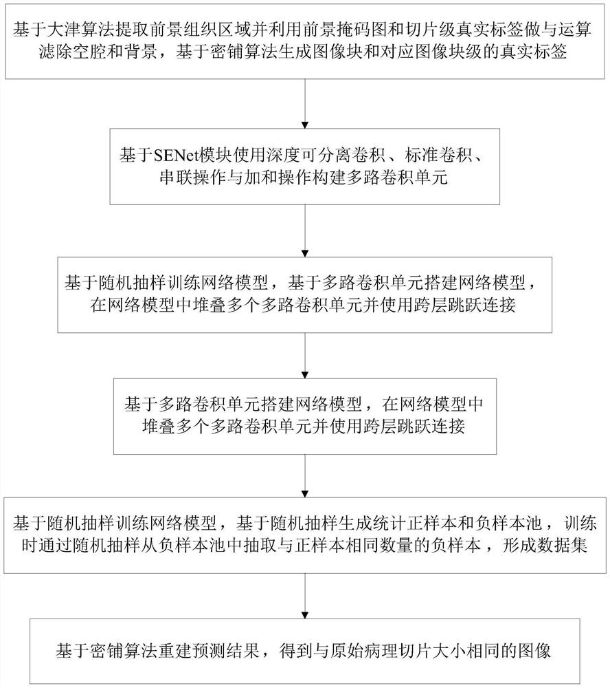
[0064] like figure 1 As shown, this embodiment provides a method for segmenting and predicting gastric cancer pathological slice images based on SENet, including the following steps:

[0065] S1: Extract the foreground tissue area based on the Otsu algorithm and use the foreground mask map and slice-level real labels to filter out cavities and backgrounds, and generate image blocks and corresponding image block-level real labels based on the dense tiling algorithm. The specific steps include:

[0066] According to the annotation file, the positive area annotation mask map is generated, the foreground tissue is extracted from the pathological slice based on the Otsu algorithm to generate the tissue mask map, and then the annotation mask map and the tissue mask map are ANDed to obtain the annotation mask for filtering out the cavity picture;

[0067] Set a square sliding window in the upper left corner of the original slice and labeling mask image, and start sliding with the wi...

Embodiment 2

[0094] This embodiment provides an SENet-based gastric cancer pathological slice image segmentation prediction system, including: an image preprocessing module, a multi-channel convolution unit construction unit, a network model construction module, a training module, and a prediction module;

[0095] In this embodiment, the image preprocessing module is used to extract the foreground tissue region based on the Otsu algorithm, filter out the cavity and the background by using the foreground mask map and the slice-level real label to do an AND operation, and generate image blocks and corresponding images based on the dense tiling algorithm. block-level ground truth labels;

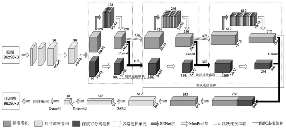
[0096] In this embodiment, the multi-channel convolution unit construction unit is used to construct a multi-channel convolution unit based on the SENet module using depthwise separable convolution, standard convolution, concatenation operation and sum operation;

[0097] In this embodiment, the network mod...

Embodiment 3

[0101] This embodiment provides a storage medium. The storage medium may be a storage medium such as a ROM, a RAM, a magnetic disk, an optical disc, etc., and the storage medium stores one or more programs. A segmentation prediction method for gastric cancer pathological slice images.


PUM


Abstract

The invention discloses a SENet-based gastric cancer pathological section image segmentation prediction method. The method comprises the following steps: generating image blocks and corresponding real labels based on an Otsu algorithm and a dense paving algorithm; a multi-path convolution unit is formed based on a SENet module; building a network model based on the multi-path convolution unit and cross-layer jump connection; training a network model based on a random sampling method; and reconstructing a prediction result based on a dense paving algorithm. According to the method, feature maps can be combined, richer and higher-dimension feature information can be obtained to represent images, small feature areas are prevented from being omitted, meanwhile, due to introduction of the SENet module, network learning can execute feature recalibration by utilizing global information, and therefore the answering ability of the whole network is enhanced, and the user experience is improved. The condition that the model is easy to ignore a small feature region in the training process is effectively avoided, and the accuracy of overall prediction is improved.

Description

technical field [0001] The invention relates to the technical field of image processing, in particular to a method for segmenting and predicting gastric cancer pathological slice images based on SENet. Background technique [0002] In recent years, deep learning has been widely used in pathological image research. At present, models based on CNN and UNet have realized the segmentation of digital pathological slices of gastric cancer, but these methods still have some problems: [0003] (1) The model complexity is high. The segmentation network contains a huge amount of parameters, and training the model requires a lot of hardware resources and time costs, resulting in low prediction efficiency; [0004] (2) The existing model has good effect on macroscopic detection of lesions, but poor detection effect on small lesions, resulting in a low overall detection rate of the model. However, the pathological sections of early malignant tumors often contain only a few cancerous ar...

Claims


Application Information

Patent Timeline
Patent Type & Authority Applications(China)
IPC IPC(8): G06T7/00G06T7/194G06N3/04G06N3/08
CPCG06T7/0012G06T7/194G06N3/08G06T2207/10056G06T2207/20081G06T2207/20084G06T2207/30092G06T2207/30096G06N3/048G06N3/045Y02A90/10
Inventor 万佳杰黄泳琳唐杰黄俊扬赖嘉兴裴贝
Owner SOUTH CHINA UNIV OF TECH
Learn More