Biological cell ultrastructure map library establishment method and system

A technology of ultrastructure and biological cells, applied in the field of image processing, can solve the problems of small observation range, limited value of information, and small selection of materials

Pending Publication Date: 2022-08-09
THE SECOND PEOPLES HOSPITAL OF SHENZHEN
View PDF0 Cites 0 Cited by
  • Summary
  • Abstract
  • Description
  • Claims
  • Application Information

AI Technical Summary

Problems solved by technology

Transmission electron microscopy has the advantages of high resolution and high specificity, but it also has the characteristics of small sample size and small observation range, which lead to great limitations
[0004] The ultrastructural images of biological cells collected by transmission electron microscopy focus on case-by-case studies, and are mainly used as a supplement to the identification ability of ordinary optical microscopes. The information carried by ultrastructural images of biological cells has limited value.

Method used


Image

  • Biological cell ultrastructure map library establishment method and system
  • Biological cell ultrastructure map library establishment method and system
  • Biological cell ultrastructure map library establishment method and system

Examples


Embodiment Construction

[0031] In order to make the purpose, technical solutions and advantages of the present application more clearly understood, the present application will be described in further detail below with reference to the accompanying drawings and embodiments. It should be understood that the specific embodiments described herein are only used to explain the present application, but not to limit the present application.

[0032] Unless otherwise defined, all technical and scientific terms used herein have the same meaning as commonly understood by one of ordinary skill in the art to which this invention belongs. The terms used herein in the description of the present invention are for the purpose of describing specific embodiments only, and are not intended to limit the present invention.

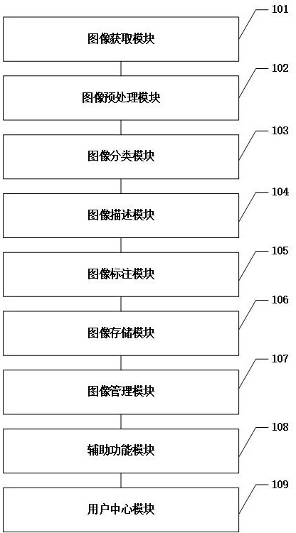
[0033] In one embodiment, the present application provides a library system of biological cell ultrastructure, such as figure 1 As shown, the system includes:

[0034] The image acquisition module ...


PUM


Abstract

The invention provides a method and system for establishing a biological cell ultrastructure gallery, and the method comprises the steps: obtaining an image collected by a biological cell through an optical microscope and an ultrastructure image collected by a transmission electron microscope which is used for preparing a sample from a same test specimen, the method comprises the steps of setting image classification, image description and image annotation rules of an image, generating a user account, distributing corresponding roles and permissions, storing the image subjected to image preprocessing, image classification, image description and image annotation processing into an image library, and realizing operations such as addition, deletion, modification and query of the image in the image library. The problem that a biological cell ultrastructure image and an optical display mirror image are logically isolated from each other and lack systematic association is solved.

Description

technical field [0001] The invention belongs to the field of image processing, and relates to a method and a system for establishing a biological cell ultrastructure library. Background technique [0002] Ultrastructural of biological cells refers to the microscopic morphology and some biological macromolecular structures in tissues and cells that cannot be distinguished by light microscopy. The resolution limit of an ordinary optical microscope is about 0.2 microns, and the thickness of the cell membrane, endoplasmic reticulum membrane and nuclear membrane, and the diameters of ribosomes, microsomes, microtubules and microfilaments are all less than 0.2 microns. To these cellular structures, to observe various submicroscopic structures in cells, electron microscopes with higher resolution must be used. Electron microscopes are mainly divided into transmission electron microscopes (Transmission Electron Microscope, TEM), scanning electron microscopes, ultra-high pressure mi...

Claims


Application Information

Patent Timeline
Patent Type & Authority Applications(China)
IPC IPC(8): G06F16/51G06F16/55G06V10/20
CPCG06F16/51G06F16/55
Inventor 王莹周玉利茹进伟
Owner THE SECOND PEOPLES HOSPITAL OF SHENZHEN
Why Eureka
Try Eureka