
System for detection and analysis of material uterine, material and fetal cardiac and fetal brain activity

A fetal heart, uterus technology, applied in the field of measurement and measurement analysis, predicting the state of a part of the body, can solve problems such as insufficient patient evaluation and diagnosis

Inactive Publication Date: 2006-12-06
REPRODIVE HEALTH TECH
View PDF0 Cites 3 Cited by
  • Summary
  • Abstract
  • Description
  • Claims
  • Application Information

AI Technical Summary

Problems solved by technology

However, the devices and methods of data acquisition and signal processing depicted in these studies appear to be insufficient for proper assessment and diagnosis of patients

Method used


Image

  • System for detection and analysis of material uterine, material and fetal cardiac and fetal brain activity
  • System for detection and analysis of material uterine, material and fetal cardiac and fetal brain activity
  • System for detection and analysis of material uterine, material and fetal cardiac and fetal brain activity

Examples


Embodiment Construction

[0048] Any one or more of the following functions, steps, frames, or components of the present invention may be included or excluded at any time during the construction or use of the instrument as required by the designer, builder, or operator, and the following Each feature or component of the invention depicted is optionally included in the invention or used by the operator of the invention.

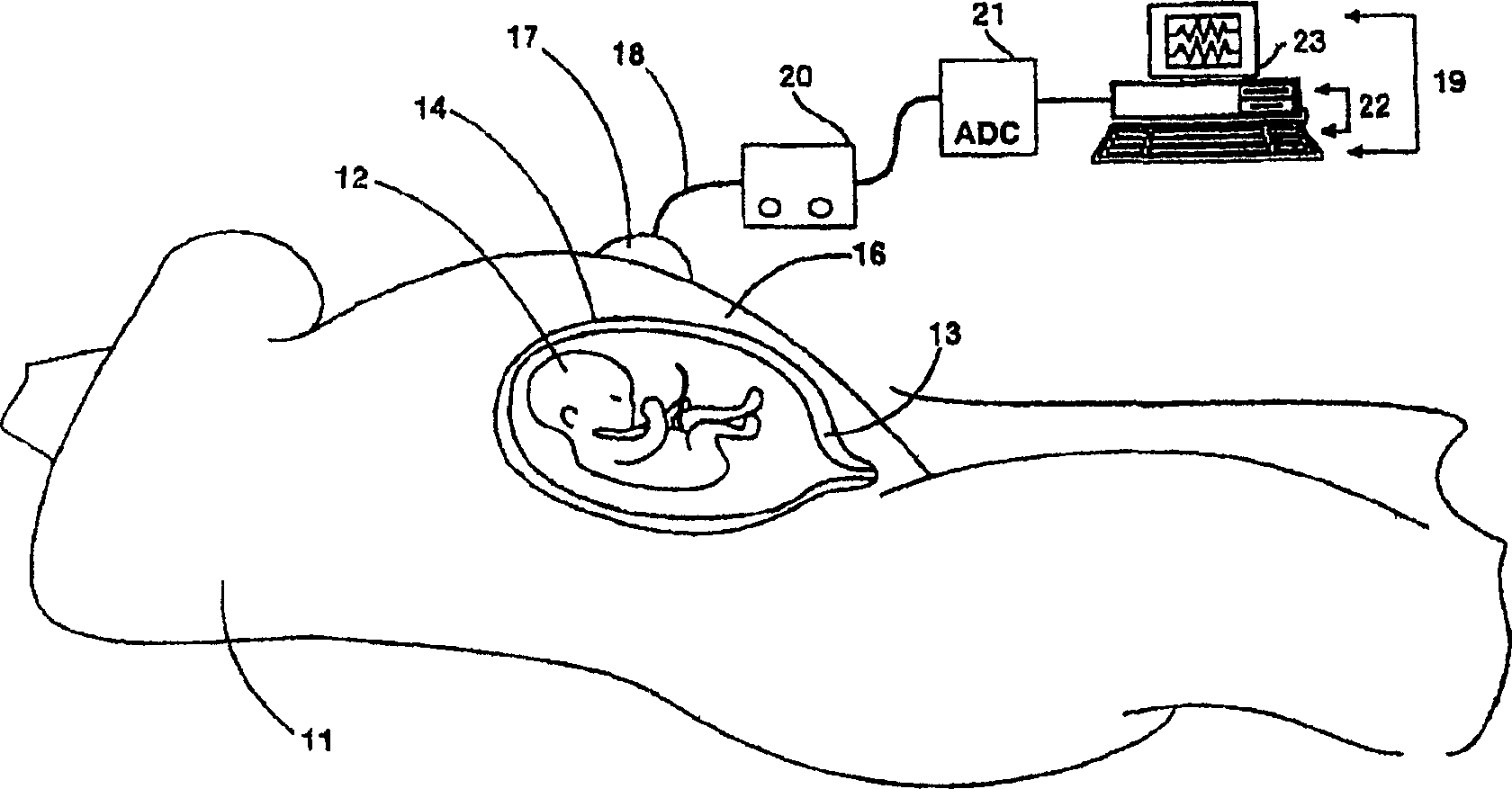
[0049] figure 1 is a schematic side view showing an internal view of a fetus in the uterus of a pregnant patient with the recording device according to the present invention attached to the surface of the abdomen of the pregnant patient. figure 1 A pregnant patient 11 is shown schematically with a fetus 12 held in a uterus 13 . The uterine wall 14 is composed primarily of muscular tissue and is located adjacent to the patient's abdominal wall 16 . In accordance with the principles of the present invention, electrodes 17 of a tripolar, quadrupolar or other multipolar configuration are...


PUM


Abstract

The present invention presents a method and apparatus for recording and analyzing uterine electrical activity, or electromyography (EMG), from the surface of the abdomen, vagina, or cervix of a patient for the purpose of diagnosing contractile patterns of the uterus in pregnant and non-pregnant women. The method and apparatus described include methods for the systematic detection, analysis, characterization and communication of infomation about electrical signals recorded from the abdominal / vaginal / cervical surface. The present invention provides data analysis techniques for analyzing the electrical data measured from the surface of a patient to characterize their uterine, abdominal, and cardiac muscle activity, as well as cardiac and brain activity of the fetus simultaneously or separately. These techniques and apparatus are appropriate for use in a clinic or through landline or wireless communication for use as a remote or home uterine / fetal monitoring system.

Description

Technical field: [0001] The present invention relates to the measurement of the electrical activity of the body. More specifically, the invention relates to measurements and their analysis to predict the state of a body part. Background technique: [0002] There is currently no objective way to evaluate uterine contractions. This is true in nonpregnant patients with severe contractions associated with dysmenorrhea, or in pregnant patients in whom the uterus sometimes moves before labor. Usually, the uterus is quiescent in nonpregnant women and during most pregnancies. At the end of pregnancy, however, the myometrium is subjected to a series of changes that result in simultaneous, rhythmic uterine contractions (labor). The diagnosis of childbirth is a very important issue facing obstetricians. In addition, preterm labor occurs in approximately 10% of pregnant patients and is difficult to diagnose. On-due or premature labor often requires adjunctive drug therapy to stimul...

Claims


Application Information

Patent Timeline
Patent Type & Authority Patents(China)
IPC IPC(8): A61B5/0488A61B5/0444A61B5/00A61B5/344
CPCA61B5/4362A61B5/0011A61B5/04085A61B5/0444A61B5/04882A61B5/7203A61B5/7225A61B5/7246A61B5/725A61B5/7257A61B5/726A61B5/4064A61B5/282A61B5/344A61B5/391
Inventor 罗伯特·E·加非尔德威廉姆·L·马耐尔
Owner REPRODIVE HEALTH TECH
Learn More