Optical projection imaging system and method for automatically detecting cells with molecular marker compartmentalization associated with malignancy and disease

A molecular marker, imaging system technology, applied in the field of projection imaging system and cell classification, can solve the problem of not being used for diagnosis, and there is no effective method.

Inactive Publication Date: 2005-08-10
VISIONGATE
View PDF5 Cites 28 Cited by
  • Summary
  • Abstract
  • Description
  • Claims
  • Application Information

AI Technical Summary

Problems solved by technology

[0012] The present invention overcomes the deficiencies of the prior art in which the measurement of subcellular and subnuclear compartmentalization of molecular markers is not useful for diagnosis because, until now, no efficient method of performing these measurements existed

Method used


Image

  • Optical projection imaging system and method for automatically detecting cells with molecular marker compartmentalization associated with malignancy and disease
  • Optical projection imaging system and method for automatically detecting cells with molecular marker compartmentalization associated with malignancy and disease
  • Optical projection imaging system and method for automatically detecting cells with molecular marker compartmentalization associated with malignancy and disease

Examples


Embodiment Construction

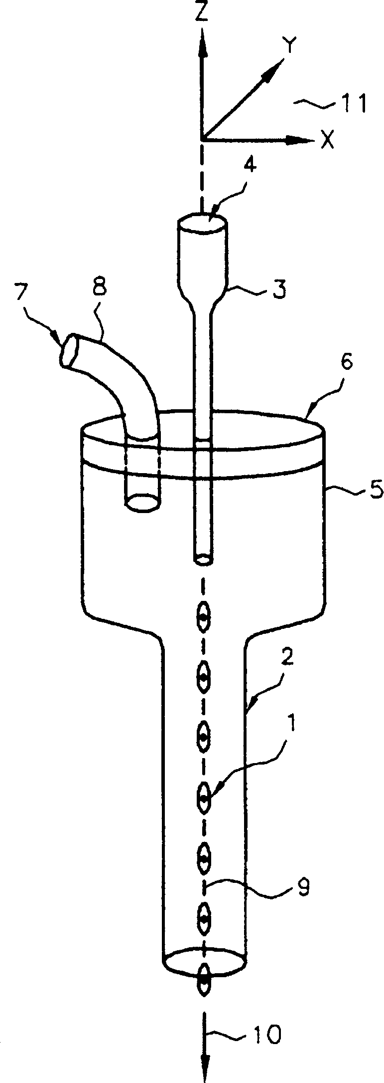
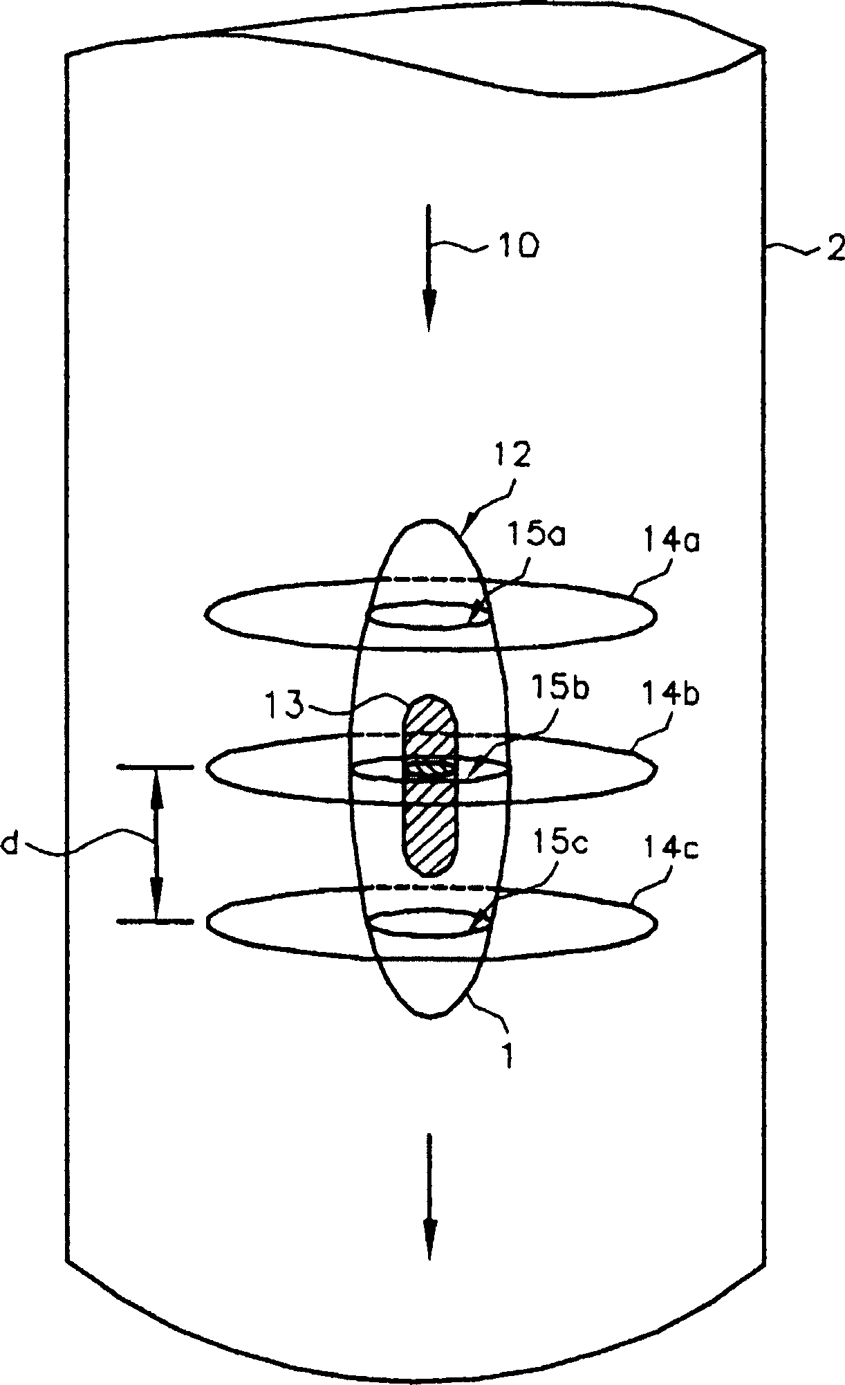
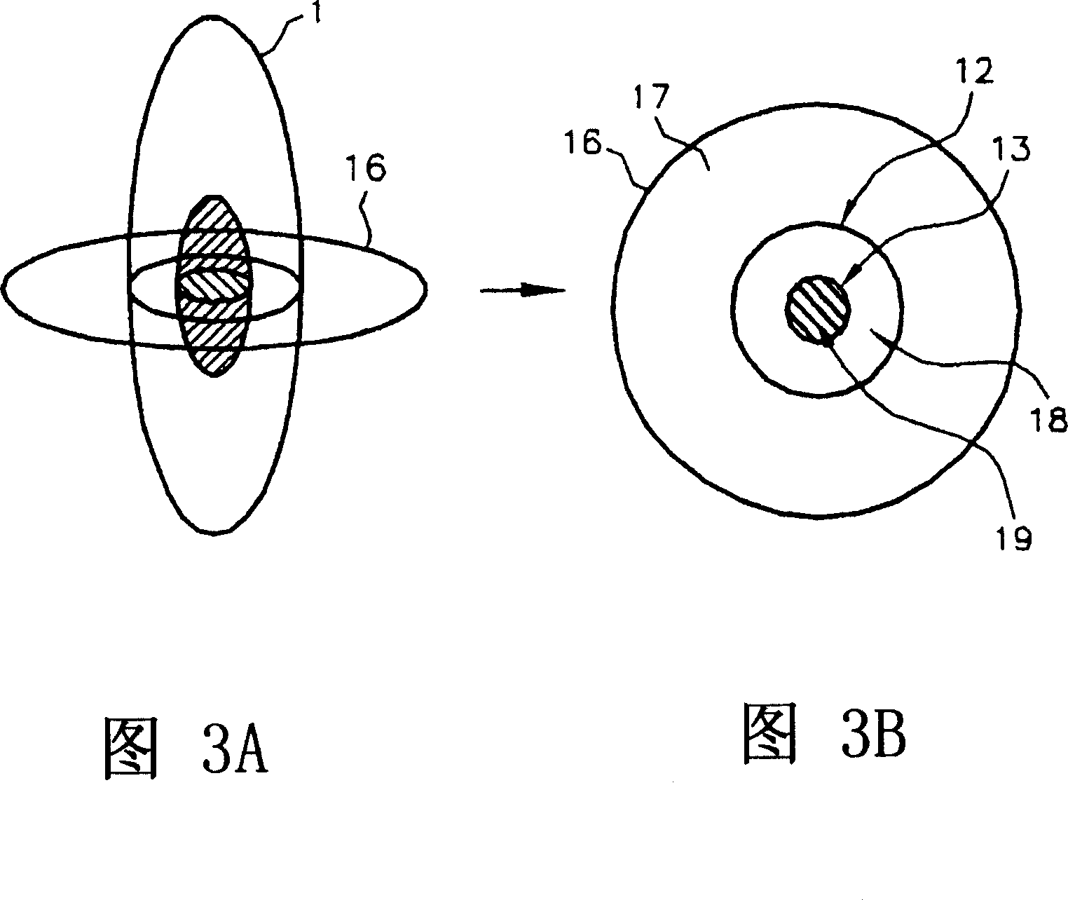
[0030] The invention is described herein with respect to specific examples involving biological cells, however, it should be understood that these examples illustrate the principles of the invention and are not intended to limit the invention. In one example, the establishment of a three-dimensional distribution of spot density and emission intensity within the microscope volume allows density and fluorescence measurements to be made anywhere within the subcellular body and the determination of structures labeled with labeled molecular probes or molecular markers. Location. By using labeled molecular probes, the number of probes attached to a specific structure or molecular marker in a microscopic object can be measured, giving a measure of the division of the marker associated with that structure. For purposes of description, an object such as a biological cell can be labeled with at least one labeled molecular probe, such as an antibody, a specific binding protein, a lectin ...


PUM

PropertyMeasurementUnit
widthaaaaaaaaaa

Abstract

Detection of cells with molecular marker compartmentalization associated with malignancy and / or disease including a cancer by obtaining a cell sample and suspending cells (1) in a solution (100). The cells are fixed (102). A molecular marker is labeled with tagged molecular probes (103). A cell (1) is illuminated with a point source of light (104), and different projection images are obtained with a digital array detector (105). The projection images are compensated for variations in illumination (106), and analyzed to detect a labeled molecular marker having densitometric and location features (107). The location of the molecular marker is analyzed (108), and compartmentalization of the molecular marker is measured using the densitometric and location features (109). Compartmentalization of the labeled molecular marker with at is analyzed using a classifier for the detection of malignancy and / or disease associated molecular marker-compartmentalization (110).

Description

[0001] related application [0002] This application is a continuation-in-part of US Patent 6,522,775 to Alan C. Nelson, published February 18, 2003, which is incorporated herein by reference. technical field [0003] The present invention relates generally to projection imaging systems and cell sorting, and more particularly to high-throughput automated systems using projection imaging, such as flow optical tomography (FOT), according to the classification of nuclear or cytoplasmic molecular markers associated with malignancy and disease. A highly quantitative measure of detection of cancer in high-risk individuals. Background technique [0004] The most common method of diagnosing cancer in a patient is by obtaining a sample of suspected tissue and examining it under a microscope for the presence of overtly malignant cells. While the procedure is relatively straightforward when the anatomical location of the suspected tissue is known and can be fully sampled (e.g., the ce...

Claims


Application Information

Patent Timeline
Patent Type & Authority Applications(China)
IPC IPC(8): G01N21/64G01N15/10G01N15/14G01N21/27G01N23/04G01N33/48G01N33/50G01N33/574G06K9/00G06T7/00G06T11/00
CPCG01N33/5091G01N2223/419G06T2207/30024G01N33/574G01N2015/1006G01N23/046G01N15/1475G06T2207/10152G06K9/00127G06T7/0012G06V20/69
Inventor 周志伟A·C·尼尔森R·W·韦伯斯特
Owner VISIONGATE
Why Eureka
Try Eureka