Time discrimination optics imaging method and equipment used for part biological tissues of animal body

A time-resolved, optical imaging technology, applied in the field of optical imaging, which can solve problems such as the limitation of the number of luminescent molecules, the inability to obtain time-resolved data, and the spatial limitation of the distribution shape.

Inactive Publication Date: 2006-02-15
新阿特艾得凡科技研究公司
View PDF4 Cites 17 Cited by
  • Summary
  • Abstract
  • Description
  • Claims
  • Application Information

AI Technical Summary

Problems solved by technology

However, this imaging device also has certain disadvantages
For example, it requires the presence of bioluminescent molecules whose in vivo distribution shape is spatially constrained, thus greatly reducing flexibil

Method used


Image

  • Time discrimination optics imaging method and equipment used for part biological tissues of animal body
  • Time discrimination optics imaging method and equipment used for part biological tissues of animal body
  • Time discrimination optics imaging method and equipment used for part biological tissues of animal body

Examples


Embodiment Construction

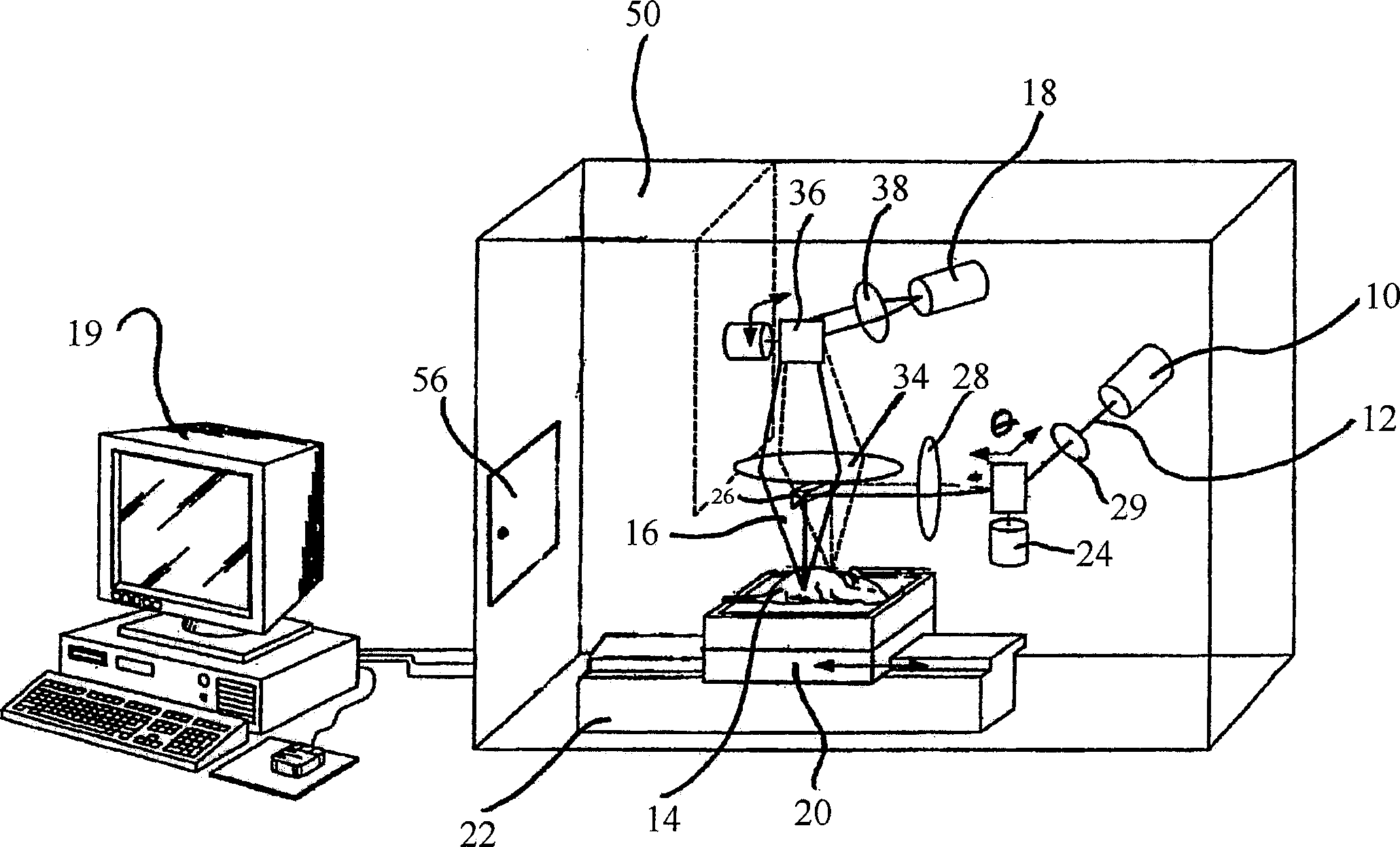
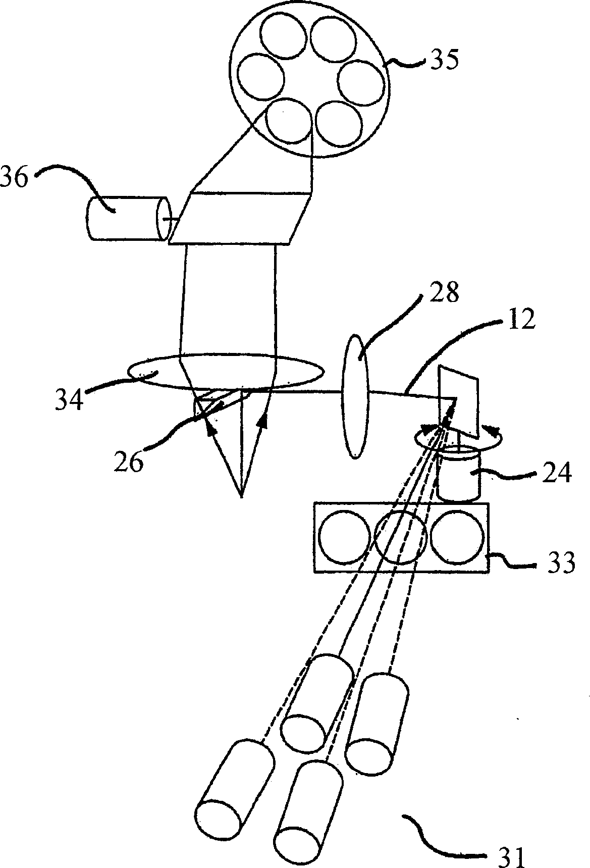
[0026] The invention relates to the field of optical imaging of turbid media such as part biological tissues of animals. Although the following description of the preferred embodiments provides examples of imaging small mammals such as mice, it should be understood that the method can also be used in larger animals, particularly laboratory animals such as dogs, pigs and primates.

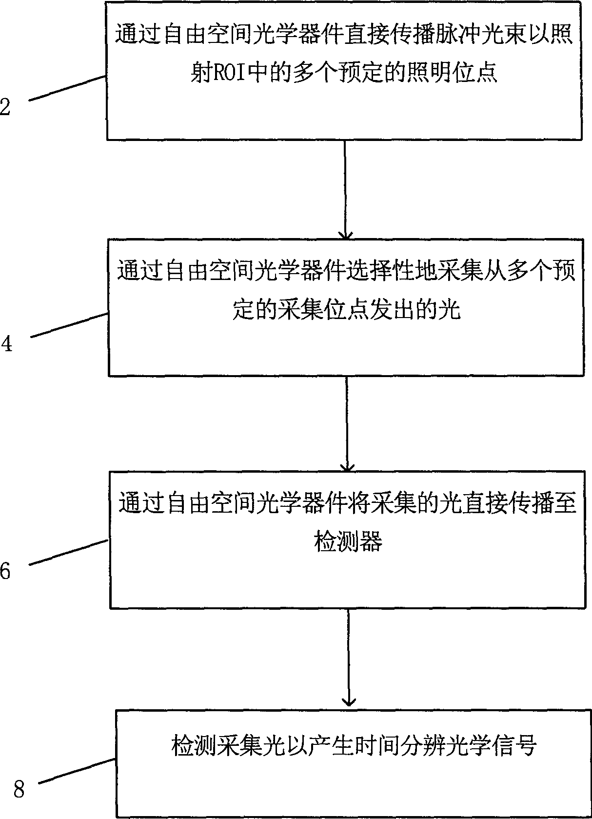
[0027] see figure 1 , generally describes an embodiment of the inventive method for acquiring optical data for use in time-resolved optical imaging. In step 2, pulsed light from a light source of selected intensity propagates directly in air (ie, through free-space optics) to illuminate a plurality of predetermined illumination sites in a ROI of biological tissue within the animal. Light emitted from multiple collection sites is selectively collected by free-space optics in step 4 after being diffusely reflected by tissue, and rebroadcasted directly to a detector through free space in step 6. Fin...


PUM


Abstract

There is provided a method and system for collecting optical data for use in time resolved optical imaging wherein pulsed light is directionally propagated through free-space optics (24, 26, 28, 29) to impinge on a plurality of illumination at the surface of a biological tissue (14) such as that comprised in small animals. Light re-emitted from the tissue is collected and directionally propagated through free space optics (34, 36, 38) towards a detector (18) to produce time resolved optical signals useful for optical image reconstructions.

Description

[0001] Cross References to Related Applications [0002] This application is a continuation of U.S. Patent Application Serial No. 10 / 665,297, filed September 22, 2003, entitled "Method and Apparatus for Time-Resolved Optical Imaging of Biological Tissue Parts of Animal Body"; and pursuant to 35 USC §119(e), requiring priority of PCT application Serial No. PCT / IB02 / 04698, filed November 11, 2002, entitled "Method and Apparatus for Time-Resolved Optical Imaging of Biological Tissue" and also claims priority to U.S. Patent Application Serial No. 10 / 665,297, filed September 22, 2003, entitled "Method and Apparatus for Time-Resolved Optical Imaging of Biological Tissue Parts of Animal Body"; and 2002 Priority to U.S. Patent Application Serial No. 60 / 505352, filed December 12, 1999, entitled "Method and Apparatus for Time-Resolved Optical Imaging of Biological Tissue." [0003] This application is related to co-assigned co-pending US Patent Application Serial No. 10 / 624902, Attorney...

Claims


Application Information

Patent Timeline
IPC IPC(8): G01N21/47G01N21/49A61B5/00G02B26/10G01N33/483A61K49/00
Inventor 威廉・F・朗伊夫・贝鲁布-洛齐埃大卫・J・霍尔劳拉・麦金托什
Owner 新阿特艾得凡科技研究公司
Why Eureka
Try Eureka