
Method of inducing kidney from pluripotent stem cells

a stem cell and kidney technology, applied in the field of induction of kidneys from pluripotent stem cells, can solve the problems of not being a general therapeutic method, increasing medical expenses, and long-term complications, and achieve the effect of effectively inducing the metanephric nephron progenitor cell

Active Publication Date: 2018-09-11
NAT UNIV CORP KUMAMOTO UNIV
View PDF5 Cites 0 Cited by
  • Summary
  • Abstract
  • Description
  • Claims
  • Application Information

AI Technical Summary

Benefits of technology

The present invention is about creating new kidneys using stem cells. We can now make the cells that form the kidneys and the filters in them, which can help with diseases and drug development. By transplanting these cells into mice, we hope they can grow into a functional kidney that produces urine.

Problems solved by technology

When the kidney function is lost, since water and a variety of toxic components are accumulated, and clouding of consciousness, dyspnea due to lung edema, or hyperpotassemia results in death, it is necessary to conduct artificial dialysis.
For anemia associated with renal failure, treatment by administering erythropoietin a few times per week is performed currently, but administration throughout the life is necessary, and this causes an increase in medical expenses.
Kidney transplantation is fundamental treatment which can completely compensate for the damaged renal function, but it could not have become a general therapeutic method due to chronic deficiency of donors.
On the other hand, artificial dialysis imposes severe dietary restriction or periodic hospital visit on patients, while since it is mere compensation for the filtration function of the kidney, it causes long term complication.
Although in vitro construction of a variety of types of tissues from pluripotent stem cells has been successful, there has still been no successful case of in vitro nephrogenesis, and establishment of methodology thereof has been waited.
This is mainly due to that details of the mechanism thereof have not been clarified due to the embryological complexity of an in vivo renal differentiation process.
Furthermore, in order that the artificially reconstructed kidney functions, essential construction of a three-dimensional structure including both the “glomerulus” and the “renal tubule”, constituting a “nephron” which is its functional unit further enhances its technical difficulty.
However, there is no report that the induction of the “metanephric nephron progenitor cell” which can reconstruct both of the glomerulus and the renal tubule from pluripotent stem cells has been succeeded.

Method used


Image

  • Method of inducing kidney from pluripotent stem cells
  • Method of inducing kidney from pluripotent stem cells
  • Method of inducing kidney from pluripotent stem cells

Examples


example 1

/ Integrina8+ / PdgfraPopulation Representing Colony-Forming Nephron Progenitors

[0128]The metanephric mesenchyme gives rise to the epithelia of glomeruli (including podocytes) and renal tubules, which constitute the major parts of the nephrons, as shown by cell fate analyses involving labeling of mesenchyme expressing the transcription factor Six2. The inventors previously proved the presence of nephron progenitors by establishing a novel colony-formation assay. When dissociated metanephric mesenchymal cells, which strongly express Sall1, were plated onto feeder cells stably expressing Wnt4, single cells formed colonies that expressed glomerular and renal tubule markers (Non-Patent Literature 9: Nishinakamura et al., 2001; Non-Patent Literature 4: Osafune et al., 2006). Therefore, the Sall1-high and Six2-positive metanephric mesenchyme represents a nephron progenitor population in the embryonic kidney.

[0129]Osr1 is another metanephric mesenchyme marker and also one of the earliest ma...

example 2

ior Intermediate Mesoderm at E9.5 Containing Colony-Forming Progenitors that Contribute to the Mesonephros

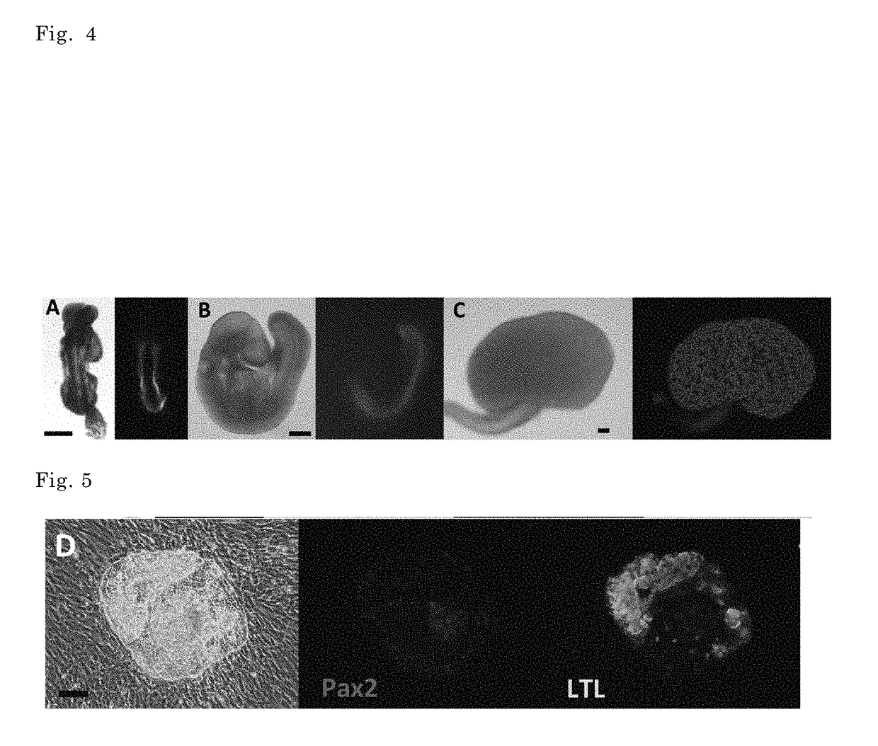
[0136]Next, the expressions of nephron progenitor markers and the colony-forming abilities of Osr1-GFP-positive cells at earlier stages were examined. As shown in FIG. 5 and Table 1, at E8.5, any overlap of GFP with Itga8 was not detected and no colonies were formed by the GFP+ population. At E9.5, colony formation by the GFP+ population was delected (0.037±0.013%).

[0137]The colony-forming cells were enriched by finding a GFP+ region that was Itga8+ / Pdgfra− (FIG. 5), and sorting of the Osr1+ / Itga8+ / Pdgfra− population (1.10±0.26%, FIG. 6; Table 2). However, even after the enrichment, the colony-forming frequency was significantly lower than those of the Osr1+ / Itga8+ / Pdgfra− populations from the metanephric region at E10.5 and E11.5 (30.9±1.5% and 50.9±5.2%, respectively; Table 2). In contrast, the colony-forming frequency of the Osr1+ / Itga8+ / Pdgfra− population from the mesonephri...

example 3

ic Nephron Progenitor Induction from the Posterior Intermediate Mesoderm at E9.5

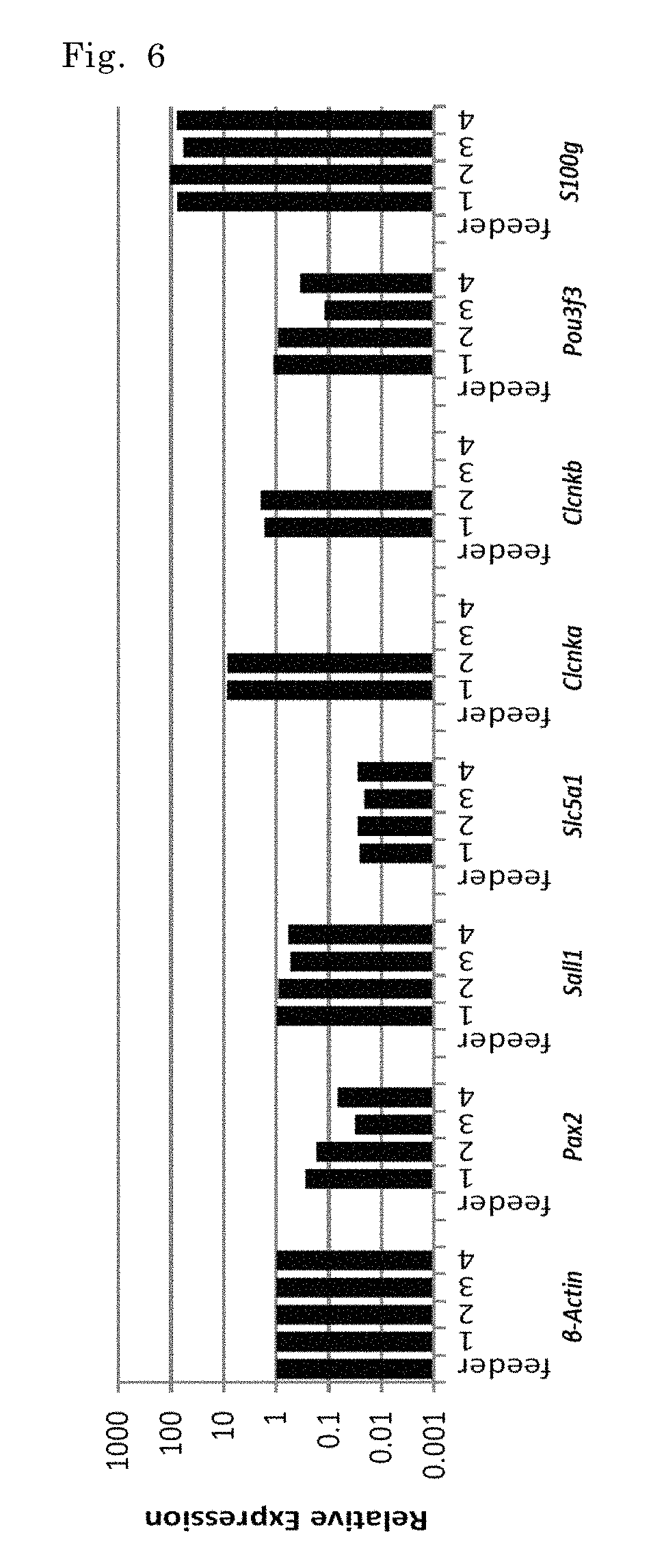
[0139]Microarray and quantitative PCR analyses were performed using the Osr1+ / Itga8+ / Pdgfra− colony-forming presumptive mesonephric progenitors at E9.5 and metanephric nephron progenitors at E10.5-E11.5. Results are shown in FIGS. 13 and 14. While both types of progenitors expressed many transcriptional factors in common, such as Osr1, Wt1,Pax2 and Six2, as well as Gdnf (a cytokine essential for kidney development), the metanephric progenitors expressed posterior Hox genes including Hoxa10, Hoxa11 and Hoxd12 more abundantly. The Hox11 family genes, which start to be expressed at the posterior end of the embryo around E9.0, are essential for metanephros development by dictating the metanephric region along the anterior-posterior axis in the intermediate mesoderm. Furthermore, a cell fate mapping study showed that the Osr1+ intermediate mesoderm at E9.5 contributes to the metanephric mesenchyme. Therefore,...


PUM

PropertyMeasurementUnit
concentrationaaaaaaaaaa

Abstract

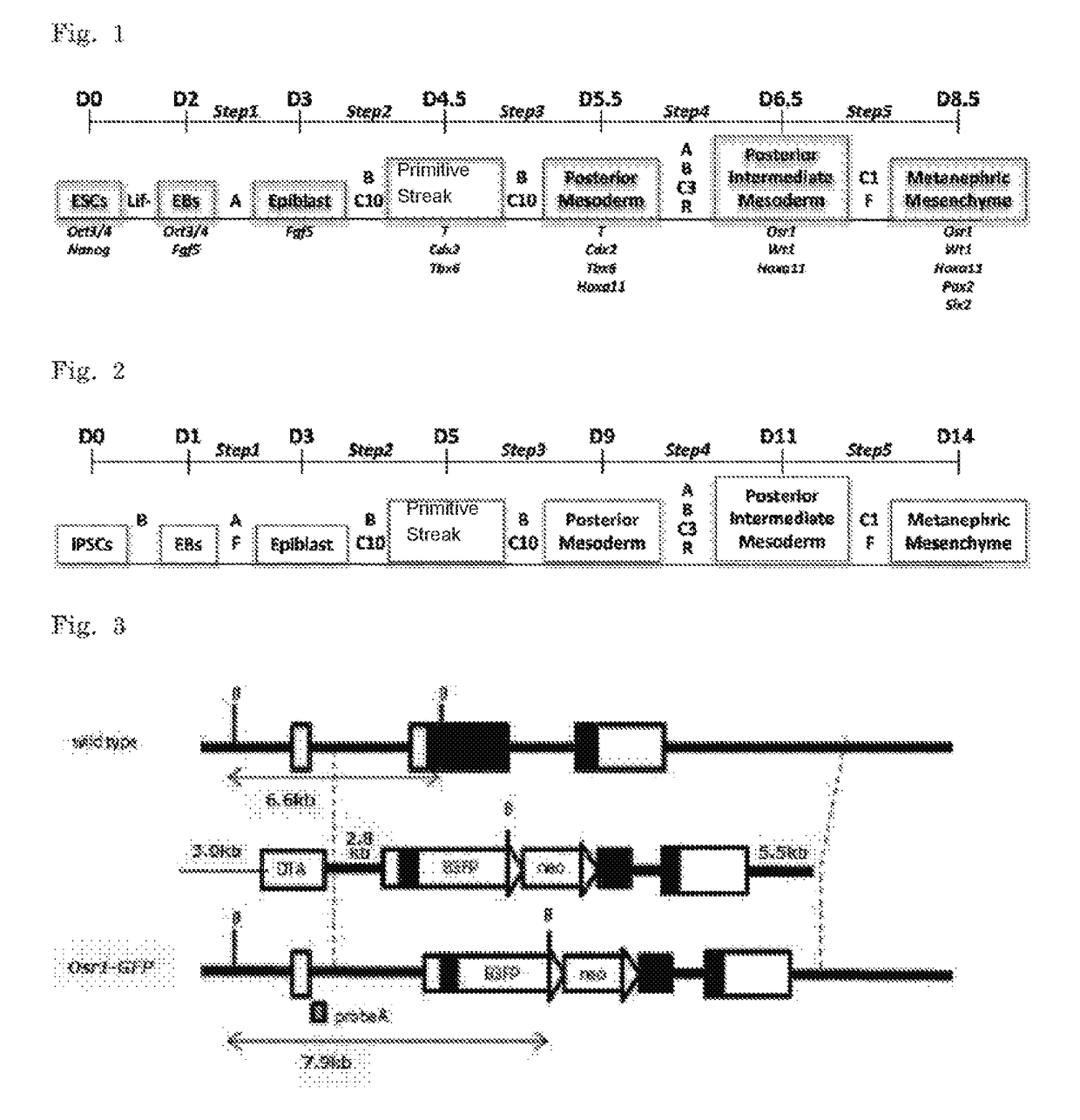
The purpose of the present invention is to provide a process or method that can be utilized when deriving a three-dimensional structure of a kidney from pluripotent stem cells such as ES cells or iPS cells. The differentiation inducing method is characterized by culturing pluripotent cells with the following three steps, in order: (a) a step of culturing an embryoid body induced from the pluripotent stem cells in medium containing Bmp4 and a high-concentration (concentration A) Wnt agonist; (b) a step of culturing the embryoid body in medium which contains activin, Bmp4, retinoic acid and a middle-concentration (concentration B) Wnt agonist; and (c) a step of culturing the embryoid body in medium containing Fgf9 and a low-concentration (concentration C) Wnt agonist (herein, the Wnt agonist concentrations is concentration A>concentration B>concentration C).

Description

CROSS-REFERENCE TO RELATED APPLICATIONS[0001]The present application is a National Stage of PCT Application No. PCT / JP2014 / 077601, filed Oct. 16, 2014, which claims priority to Japanese Application No. JP 2013-217029 filed Oct. 18, 2013. The entirety of the aforementioned applications is incorporated herein by reference.TECHNICAL FIELD[0002]The present invention relates to a method of inducing a kidney from a pluripotent stem cell. More particularly, the present invention relates to a method of inducing a three-dimensional structure of the kidney including both of a glomerulus and a renal tubule, from a pluripotent stem cell, for example, an ES cell and an iPS cell.BACKGROUND ART[0003]The kidney excretes waste products by producing urine and, at the same time, plays an important role in maintaining homeostasis of electrolytes and water in a body. When the kidney function is lost, since water and a variety of toxic components are accumulated, and clouding of consciousness, dyspnea du...

Claims


Application Information

Patent Timeline
Patent Type & Authority Patents(United States)
IPC IPC(8): C12N5/00A01N61/00C07K1/00C07K14/50C12N5/071C07K14/51
CPCC12N5/0671C12N5/0672C12N2501/119C12N2501/155C12N2506/45C12N2501/385C12N2501/415C12N2506/02C12N2501/16
Inventor NISHINAKAMURA, RYUICHITAGUCHI, ATSUHIRO
Owner NAT UNIV CORP KUMAMOTO UNIV
Learn More