
In vitro expansion of erythroid cells

a technology of erythroid cells and erythrocytes, which is applied in the field of in vitro expansion of erythroid cells, can solve the problems of increasing the amount of blood preparations used, difficulty in surgery and patient treatment, and shortage of transfusable blood supply, so as to facilitate the expansion of the cell culture scale and improve the in vitro productivity of erythrocytes

Active Publication Date: 2019-04-30
IUCF HYU (IND UNIV COOP FOUND HANYANG UNIV)
View PDF12 Cites 4 Cited by
  • Summary
  • Abstract
  • Description
  • Claims
  • Application Information

AI Technical Summary

Benefits of technology

[0032]The features and advantages of the present invention are summarized as follows:
[0033](a) the present invention using a porous structure for 3-dimensional packed cell culture of erythroid cells is effective in in vitro expansion of erythroid cells;
[0034](b) the present invention can markedly improve the in vitro productivity of erythrocytes from erythroid progenitor cells;
[0035](c) the present invention facilitates expansion of the packed cell culture scale of erythroid cells and is effective in maintaining the packed cell culture scale of erythroid cells through the pores of the microcarrier; and
[0036](d) the present invention enables the production of clinical grade erythrocytes.

Problems solved by technology

Recent rapid aging of the population and increasing demand for medical services have led to a supply shortage of transfusable blood and an increase in the amount of blood preparations used.
Thus, the current worldwide shortage of transfusable blood causes difficulties in the surgery and treatment of patients.
This blood shortage will become more serious in the future.
The risk of transfusion-transmitted infections causes serious problems in blood recipients.
Enormous costs are incurred in developing examination methods and systems to detect the infection risk but it is impossible to completely detect the infections [1].
That is, animal cell culture methods designed for cultivation conditions in existing methods and systems, such as static culture, suspension culture, fixed bed reactors, and airlift reactors, are practically impossible to apply to the culture of erythrocytes that requires an astronomical number of cells and media and spaces large enough to accommodate the cells.

Method used


Image

  • In vitro expansion of erythroid cells
  • In vitro expansion of erythroid cells
  • In vitro expansion of erythroid cells

Examples


example 1

Cell Culture and Enumeration

[0061]CD34+ cells were isolated from the cord blood of healthy donors by using the immunomagnetic microbead selection method and EasySep CD34 isolation kit (StemCell Technologies). Some of the CD34+ cells were used for culture immediately after isolation or were frozen and stored until use. The cells were cultured under stroma- and serum-free conditions for 17 days [3, 4].

[0062]Several cytokines were added to induce the CD34+ cells to differentiate and proliferate. From day 0 to day 7, the cells were cultured in media supplemented with 10 μM hydrocortisone (Sigma), 100 ng / ml stem cell factor (SCF; R&D Systems), 10 ng / ml interleukin (IL)-3 (R&D Systems), and 6 IU / ml erythropoietin (EPO; Calbiochem). From day 7 to day 13, basophilic erythroblasts were cultured in media supplemented with 50 ng / ml SCF, 10 ng / ml IL-3, and 3 IU / ml EPO. From day 13 to day 17, 2 IU / ml EPO was added [3, 4]. On day 17, when the proportions of polychromatic erythroblasts and orthoch...

example 2

Imaging of Erythroid Cells

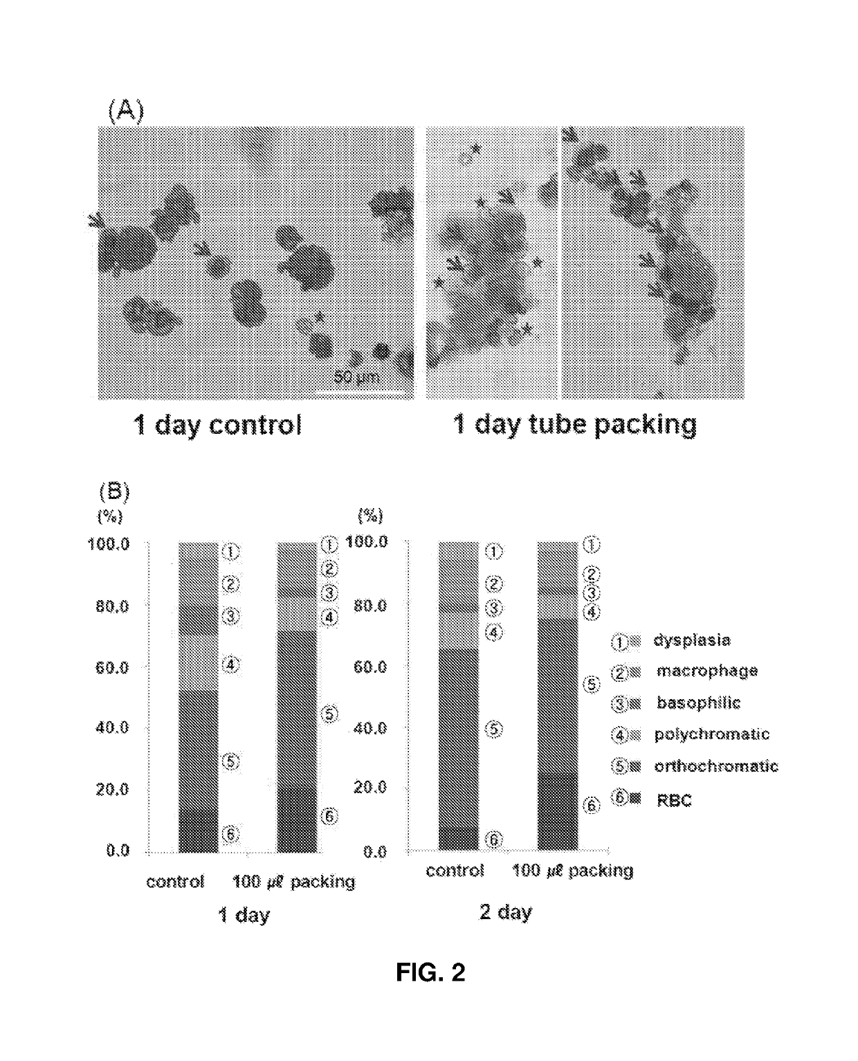
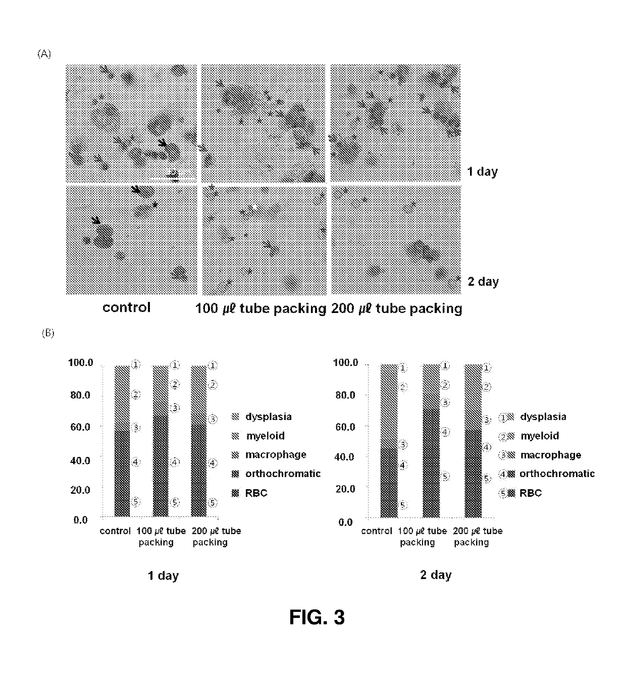
[0064]Cells were plated on a slide using a cell centrifuge (Cellspin, Hanil Science Industrial) and subsequent Wright-Giemsa staining (Sigma-Aldrich). The stained cells were analyzed for maturation stage, myelodysplasia, and cell integrity.

example 3

2D Plate Culture and 3D Packed Cell Culture

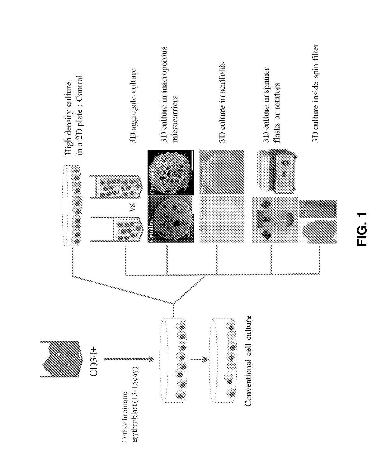
[0065]On days 15-17 of culture, the erythroid cells (polychromatic / orthochromatic erythroblasts) at the terminal maturation stage were divided and cultured under the conditions shown in FIG. 1. The control cells were cultured at a density of 1×10⁶ cells / mL in a 6-well 2D plate. In the experimental groups, the cells were allowed to be packed at a density of 2×10⁷ cells / mL in tubes and cultured in a state in which the tubes stood upright. Basal media used in the control and the experimental groups were supplemented with 5% plasma-derived serum [5]. After culture for 24 and 48 h, the cell numbers and viabilities were determined and the maturation stages of cells were confirmed using Wright-Giemsa staining.


PUM

PropertyMeasurementUnit
size distributionaaaaaaaaaa
sizeaaaaaaaaaa
volumeaaaaaaaaaa

Abstract

The present invention relates to a method for in vitro expansion of erythroid cells. The method includes subjecting erythroid cells to 3-dimensional packed cell culture using a porous structure. The use of the composition according to the present invention enables in vitro expansion of erythroid cells in the most efficient manner.

Description

CROSS-REFERENCE TO RELATED APPLICATIONS[0001]This application is a national phase application of PCT / KR2015 / 003483, filed on Apr. 7, 2015, which claims the benefit of priority to Korean Patent Application No. 10-2014-0041320, filed on Apr. 7, 2014, the disclosure of which is incorporated herein by reference.FIELD[0002]The present invention was supported by the Ministry of Health and Welfare of the Republic of Korea under Project No. HI10C17400200, which was conducted in the research project entitled “Advanced medical technology development” within the project named “Research on efficient differentiation and mechanism of stem cells into erythroid progenitor cells” by the Industry-Academic Cooperation Foundation, Yonsei University under the management of the Korea Health Industry Development Institute, from Apr. 1, 2013 to Mar. 31, 2014.[0003]The present invention was also supported by the Ministry of Health and Welfare of Republic of Korea under Project No. HI12C0202, which was condu...

Claims


Application Information

Patent Timeline
Patent Type & Authority Patents(United States)
IPC IPC(8): C12N5/00C12N5/078
CPCC12N5/0641C12N2501/125C12N2501/14C12N2501/2303C12N2531/00C12N2502/1311C12N2502/1323C12N2502/1358C12N2521/00C12N2501/39
Inventor BAEK, EUN JUNGLEE, EUN-MI
Owner IUCF HYU (IND UNIV COOP FOUND HANYANG UNIV)
Learn More