Two-stage method for treating calcified lesions within the wall of a blood vessel

a blood vessel wall and calcification technology, applied in the field of treatment system for percutaneous coronary angioplasty or peripheral angioplasty, can solve the problems of rebound, dissection, thrombosis, undue stress on the free wall of the blood vessel,

Active Publication Date: 2020-07-07
SHOCKWAVE MEDICAL
View PDF266 Cites 18 Cited by
  • Summary
  • Abstract
  • Description
  • Claims
  • Application Information

AI Technical Summary

Problems solved by technology

With such pressures comes trauma to the vessel wall which can contribute to vessel rebound, dissection, thrombus formation, and a high level of restenosis.
Non-concentric calcified lesions can result in undue stress to the free wall of the vessel when exposed to high pressures.
That energy is then released and results in the rapid expansion of the balloon to its maximum dimension and may stress and injure the vessel walls.

Method used


Image

  • Two-stage method for treating calcified lesions within the wall of a blood vessel
  • Two-stage method for treating calcified lesions within the wall of a blood vessel
  • Two-stage method for treating calcified lesions within the wall of a blood vessel

Examples


Embodiment Construction

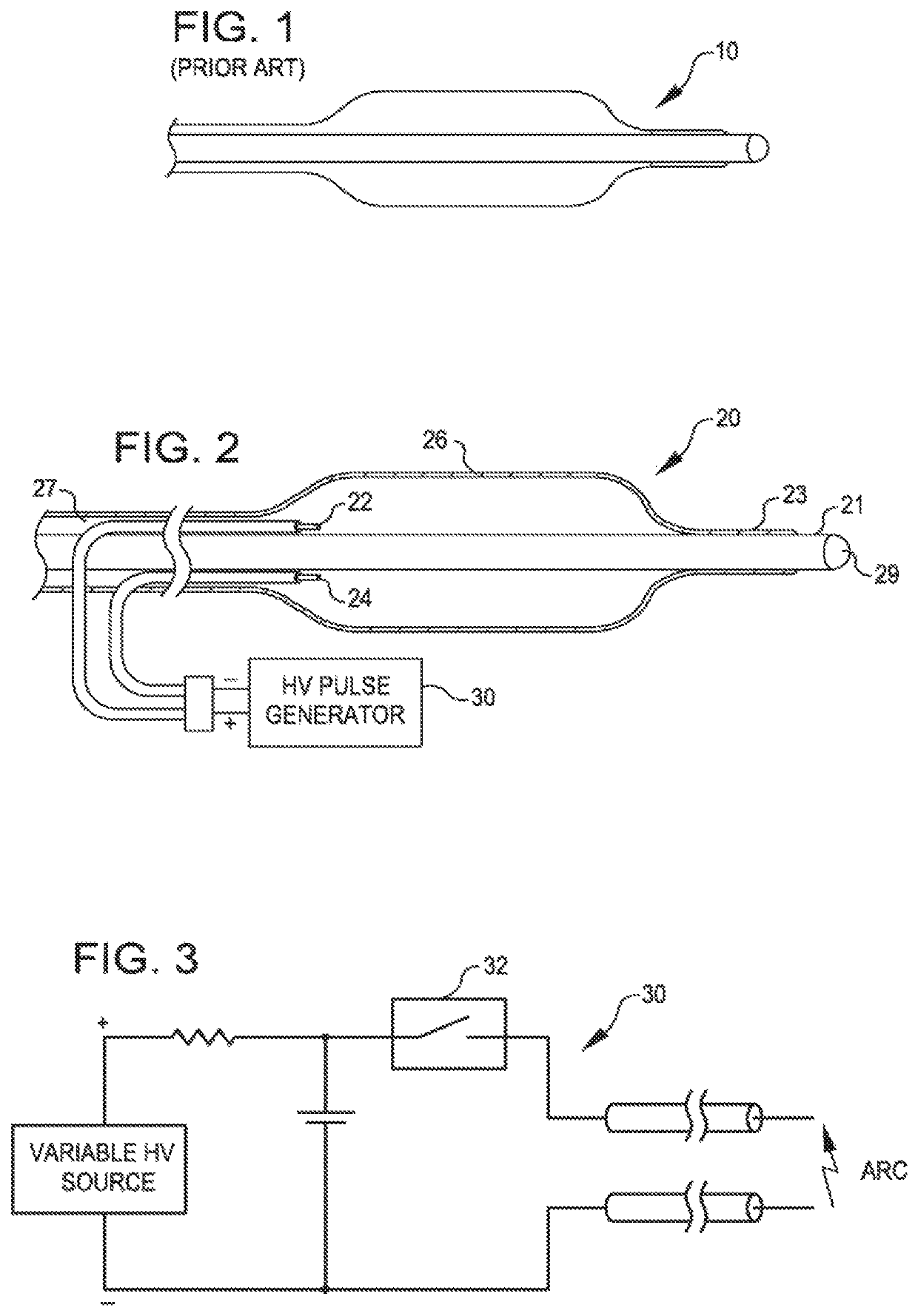
[0051]FIG. 1 is a view of the therapeutic end of a typical prior art over-the-wire angioplasty balloon catheter 10. Such catheters are usually non-complaint with a fixed maximum dimension when expanded with a fluid such as saline.

[0052]FIG. 2 is a view of a dilating angioplasty balloon catheter 20 according to an embodiment of the invention. The catheter 20 includes an elongated carrier, such as a hollow sheath 21, and a dilating balloon 26 formed about the sheath 21 in sealed relation thereto at a seal 23. The balloon 26 forms an annular channel 27 about the sheath 21 through which fluid, such as saline, may be admitted into the balloon to inflate the balloon. The channel 27 further permits the balloon 26 to be provided with two electrodes 22 and 24 within the fluid filled balloon 26. The electrodes 22 and 24 are attached to a source of high voltage pulses 30. The electrodes 22 and 24 are formed of metal, such as stainless steel, and are placed a controlled distance apart to allow ...


PUM


Abstract

A method is disclosed for treating calcified lesions within a wall of a blood vessel. The first step includes breaking apart a calcified lesion using a plurality of shockwaves generated in an angioplasty balloon of an angioplasty catheter device. The angioplasty balloon is dilated via a fluid to a first extent to fit against at least a portion of the wall of the blood vessel. A plurality of electrical pulses are delivered to a pair of electrodes disposed within the fluid inside the balloon. The electrical pulses have an amplitude sufficient to create plasma arcs in the fluid to generate shockwaves that are conducted through the fluid and through the balloon to the blood vessel, to crack the calcified lesion. After breaking apart the calcified lesion, the angioplasty balloon is allowed to further expand to a second extent greater than the first extent, thereby expanding an opening in the blood vessel.

Description

PRIORITY CLAIM[0001]The present application is a continuation-in-part of U.S. patent application Ser. No. 16 / 028,225, filed Jul. 5, 2018, which is in turn a continuation of U.S. patent application Ser. No. 14 / 660,539, filed Mar. 17, 2015, now issued as U.S. Pat. No. 10,039,561 on Aug. 7, 2018, which is a continuation of U.S. patent application Ser. No. 13 / 646,570, filed Oct. 5, 2012, now issued as U.S. Pat. No. 9,011,462, on Apr. 21, 2015, which is a continuation of U.S. patent application Ser. No. 12 / 482,995, filed Jun. 11, 2009, now issued as U.S. Pat. No. 8,956,371 on Feb. 17, 2015, which_claims the benefit of U.S. Provisional Patent Application Ser. No. 61 / 061,170, filed Jun. 13, 2008, each of which is incorporated herein by reference in its entirety.BACKGROUND OF THE INVENTION[0002]The present invention relates to a treatment system for percutaneous coronary angioplasty or peripheral angioplasty in which a dilation catheter is used to cross a lesion in order to dilate the lesio...

Claims


Application Information

Patent Timeline
Patent Type & Authority Patents(United States)
IPC IPC(8): A61B17/22A61B17/225A61B17/3207
CPCA61B17/2202A61B17/22029A61B17/22022A61B2017/22024A61B17/320725A61B17/2251A61B2017/22021A61B2017/22038A61B2017/22058A61B2017/22061A61B2017/22001A61B2017/22025A61B2017/22062
Inventor ADAMS, JOHN M.HAWKINS, DANIELALFERNESS, CLIFTON A.
Owner SHOCKWAVE MEDICAL
Why Eureka
Try Eureka