Method and apparatus for reconstructing a three-dimensional representation of a target volume inside an animal or human body

a three-dimensional representation and target volume technology, applied in image data processing, diagnostics, applications, etc., can solve the problems of unnecessary longness, less accurate diagnosis and subsequent treatment of patients, and achieve the effect of accurate diagnosis and subsequent treatment, discomfort, and beneficial radiation exposure tim

Active Publication Date: 2020-08-11
MEDIS ASSOC
View PDF4 Cites 26 Cited by
  • Summary
  • Abstract
  • Description
  • Claims
  • Application Information

AI Technical Summary

Benefits of technology

[0007]It is an object of the invention to provide an improved method and apparatus for reconstructing a three-dimensional representation of a target volume inside an animal or human body allowing for a faster and more sophisticated imaging technique, thus reducing the acquisition and the analyzing time of the imaging diagnosis and limiting the imaging treatment session for the patient as well as avoiding diagnosis errors by the diagnostician.
[0010]Accordingly, with these features an improved reconstructing method is obtained, wherein an optimal set of two-dimensional image projections is obtained and proposed, based on which a correct three-dimensional contour image of said target volume is being reconstructed. Thus with this correct three-dimensional contour image the diagnostician is capable of performing a more accurate diagnosis and subsequent treatment of the patient, which in turn is subjected to a less lengthy imaging treatment session, which is beneficial in terms of radiation exposure time, discomfort, etc.
[0013]By defining a first subset of second operational orientation settings of said imaging means for orientation in said second imaging position, wherein said first subset of second operational orientation settings is distinct from said first operational orientation setting any duplication of acquiring identical or more or less identical two-dimensional image projections is being avoided. Herewith the length if the imaging treatment session is further reduced (which is beneficial for the patient), but also incorrect or unsuitable three-dimensional contour images are not reconstructed thus avoiding incorrect analysis and diagnosis of the target volume of the patient by the diagnostician.
[0021]In particular said first subset of second operational orientation settings limit an angle between a first plane in which said first imaging position corresponding to said first operational orientation setting is defined and a second plane in which said second imaging positions corresponding to said second operational orientation settings are defined between a range of 35°-50°. Herewith the acquisition of an incorrect set of two-dimensional image projections and an inaccurate three-dimensional contour image based on this set is thus avoided further improving the reconstructing method according to the invention.
[0025]Accordingly, with these features an improved apparatus for reconstructing a three-dimensional representation of a target volume is obtained, wherein an optimal set of two-dimensional image projections is obtained and proposed, based on which a correct three-dimensional contour image of said target volume is being reconstructed. Thus with this correct three-dimensional contour image the diagnostician is capable of performing a more accurate diagnosis and subsequent treatment of the patient, which in turn is subjected to a less lengthy imaging treatment session, which is beneficial in terms of radiation exposure time, discomfort, etc.

Problems solved by technology

This tends to lead to unnecessary lengthy imaging treatment session for the patient, which is undesirable due to radiation exposure, but also to incorrect selection of the correct set of image projections resulting in less accurate diagnosis and subsequent treatment of the patient.

Method used


Image

  • Method and apparatus for reconstructing a three-dimensional representation of a target volume inside an animal or human body
  • Method and apparatus for reconstructing a three-dimensional representation of a target volume inside an animal or human body
  • Method and apparatus for reconstructing a three-dimensional representation of a target volume inside an animal or human body

Examples


Embodiment Construction

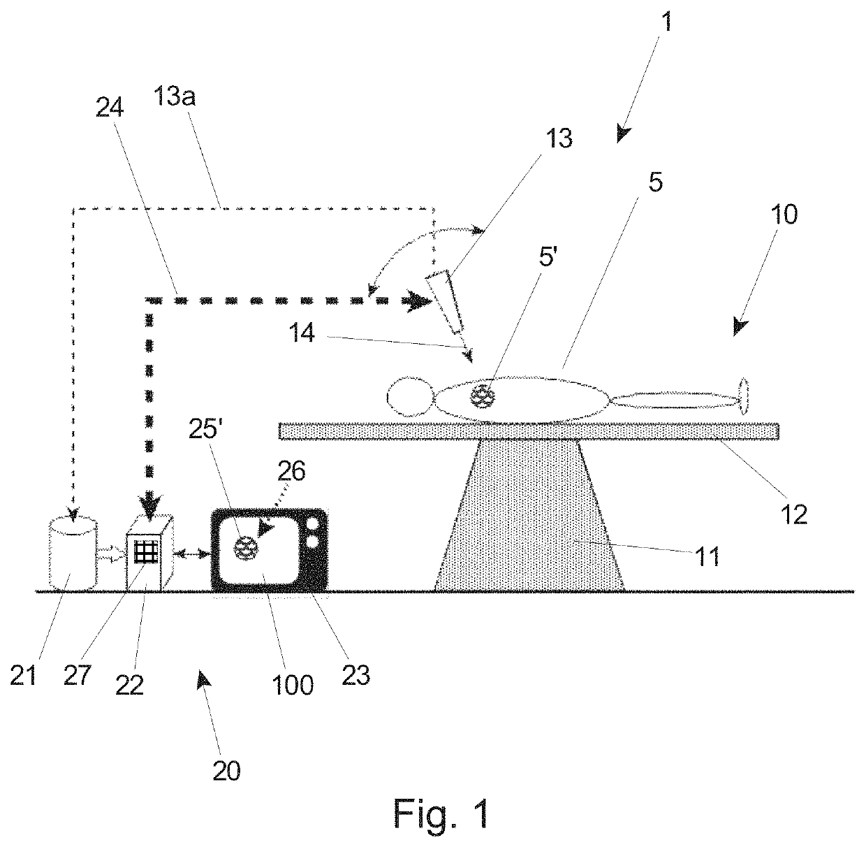
[0031]FIG. 1 shows in very schematic form various elements of a known imaging device 10. An object 5, here a human patient 5 is shown lying in lithotomy position on a table 12 which is supported by a base 11. Above the table 12 imaging means 13 are positioned which imaging means 13 are movable accommodated in a structure (not shown) forming part of the imaging device 10. This allows for a controlled positioning of the imaging means 13 at any angle or orientation relative to a target volume 5′ inside the patient's body 5.

[0032]The imaging means 13 can be any imaging technique capable of obtaining 2D image projection of a target volume (here indicate with reference numeral 5) in an object 5. For example X-ray radiation imaging means can be used when implementing the method and apparatus according to the invention.

[0033]It should be noted that the object 5 lying on the table 12 can be a human or animal body or any other object which is to be subjected to the apparatus and method accord...


PUM


Abstract

The invention relates to method and apparatus for reconstructing a three-dimensional representation of a target volume inside an animal or human body.Hereto, according to the method step ii) is preceded by the steps of:a1) displaying said first two-dimensional image projection being obtained in step i) for a user using said displaying means,a2) acquiring a first operational orientation setting of said imaging means corresponding with said first imaging position;a3) defining based on said first operational orientation setting a first set of second operational orientation settings of said imaging means for orientation in a second imaging position;a4) selecting from said set one of said second operational orientation settings;a5) using said second operational orientation setting being selected for acquiring said second two-dimensional image projection of said target volume using said imaging means being positioned in the second imaging position corresponding to said second operational orientation setting.Accordingly, with these features an improved reconstructing method is obtained, wherein an optimal set of two-dimensional image projections is obtained and proposed, based on which a correct three-dimensional contour image of said target volume is being reconstructed. Thus with this correct three-dimensional contour image the diagnostician is capable of performing a more accurate diagnosis and subsequent treatment of the patient, which in turn is subjected to a less lengthy imaging treatment session, which is beneficial in terms of radiation exposure time, discomfort, etc.

Description

TECHNICAL FIELD AND BACKGROUND[0001]The invention relates to method for reconstructing a three-dimensional representation of a target volume inside an animal or human body, said method comprising the steps of:i) acquiring a first two-dimensional image projection of said target volume using imaging means being positioned in a first imaging position,ii) acquiring a second two-dimensional image projection of said target volume using said imaging means being positioned in a second imaging position,iii) reconstructing from said two-dimensional image projections a three-dimensional contour image of said target volume using transformation means,iv) displaying said three-dimensional contour image of said target volume fora user using displaying means.[0002]The invention also relates to an apparatus for reconstructing a three-dimensional representation of a target volume inside an animal or human body, the apparatus comprising: an imaging means configured to obtain at least a first and a sec...

Claims


Application Information

Patent Timeline
Patent Type & Authority Patents(United States)
IPC IPC(8): G06T17/00A61B6/00G06T11/00A61B6/02
CPCA61B6/025G06T11/003G06T11/008A61B6/545A61B6/508A61B6/504A61B6/466G06T17/00A61B6/027A61B6/5205A61B6/547A61B6/486
Inventor REIBER, JOHAN HENDRIKUS CHRISTIAANJANSSEN, JOHANNES PETRUSKONING, GERHARDTU, SHENGXIAN
Owner MEDIS ASSOC
Why Eureka
Try Eureka