Extracellular vesicle isolation method using metal

a technology of extracellular vesicles and metals, applied in the field of metal isolating extracellular vesicles, can solve the problems of large amount of samples required, excessive amount of labor and time, and large amount of labor and time, and achieve rapid and effective isolation of extracellular vesicles, maximize the efficiency of extracellular vesicles, and efficient isolating extracellular vesicles

Active Publication Date: 2021-10-05
ROSETTA EXOSOME
View PDF15 Cites 1 Cited by
  • Summary
  • Abstract
  • Description
  • Claims
  • Application Information

AI Technical Summary

Benefits of technology

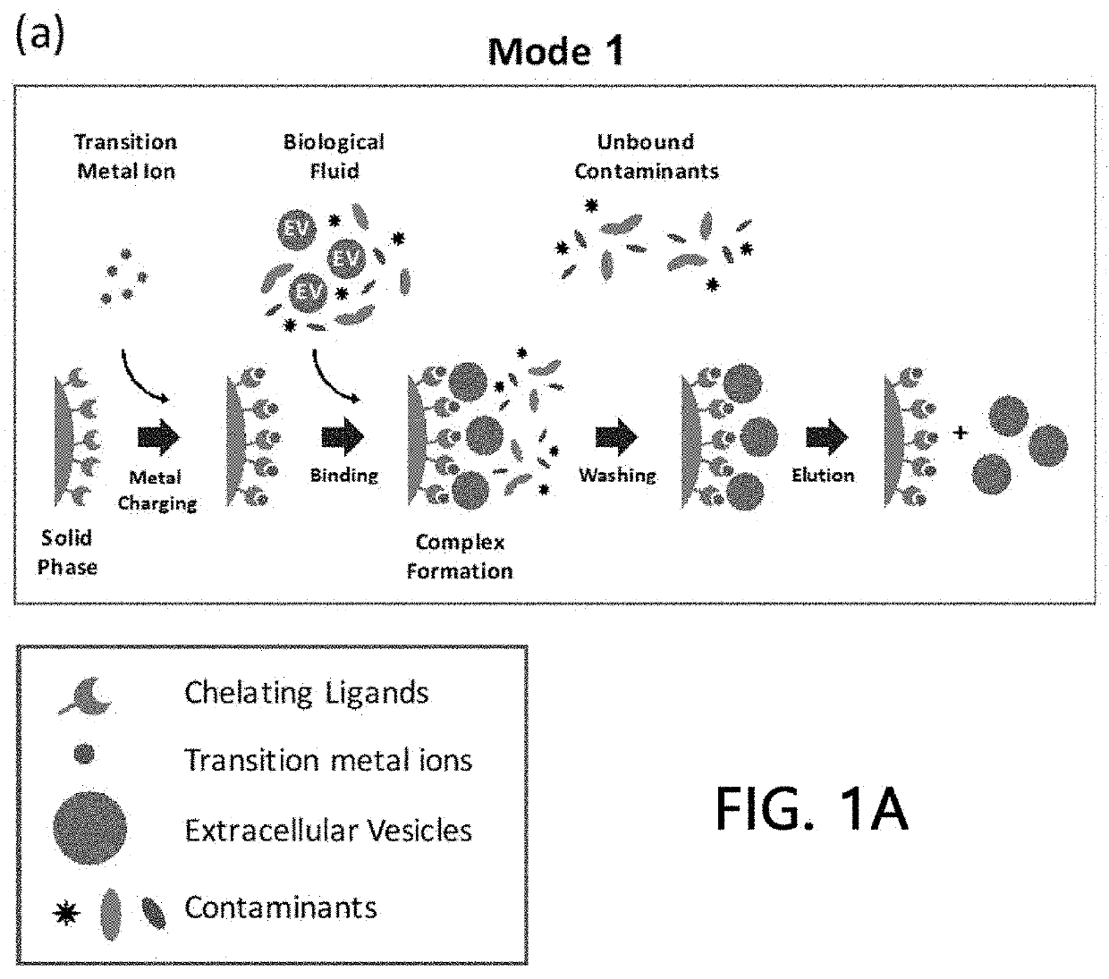
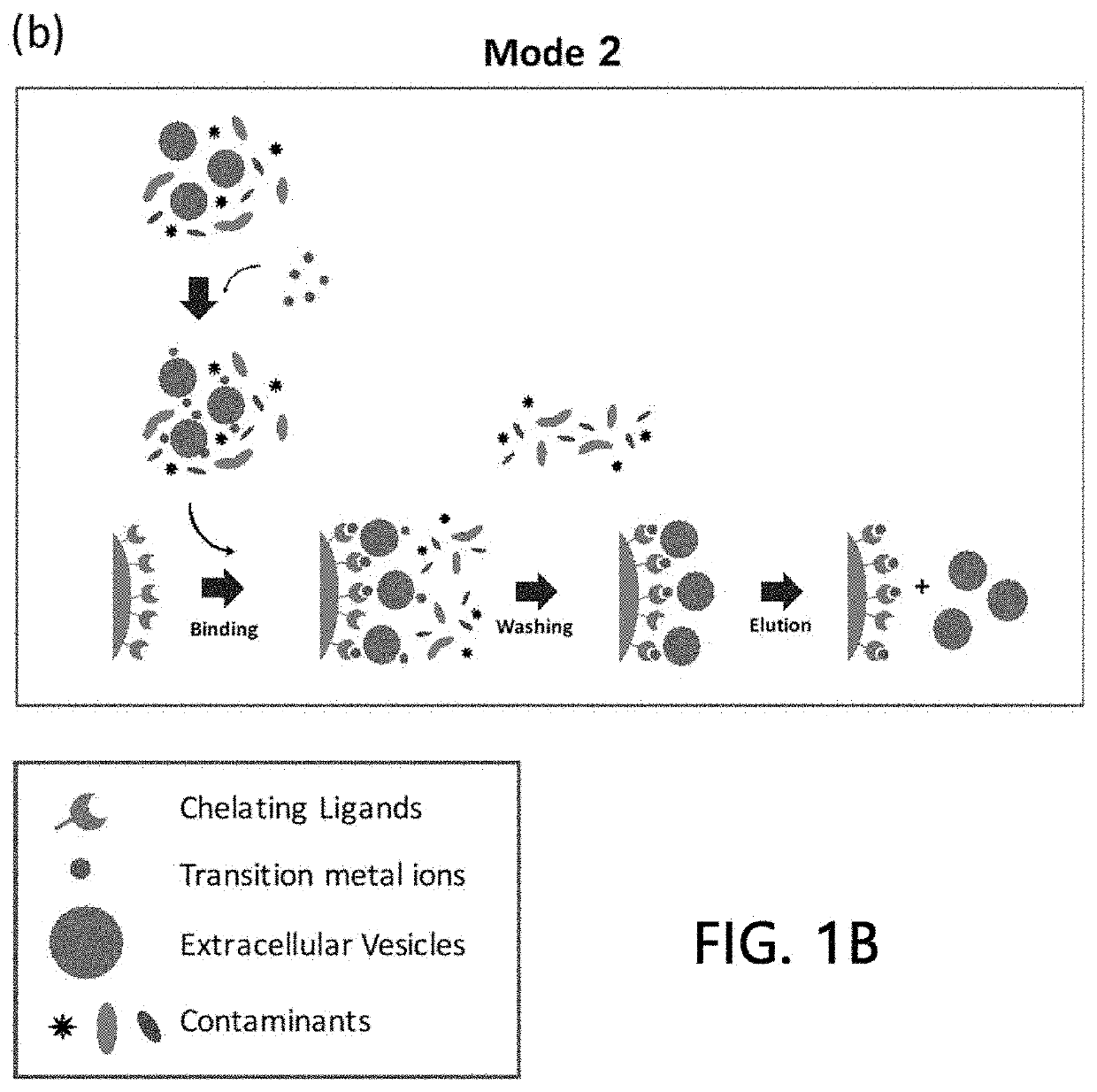
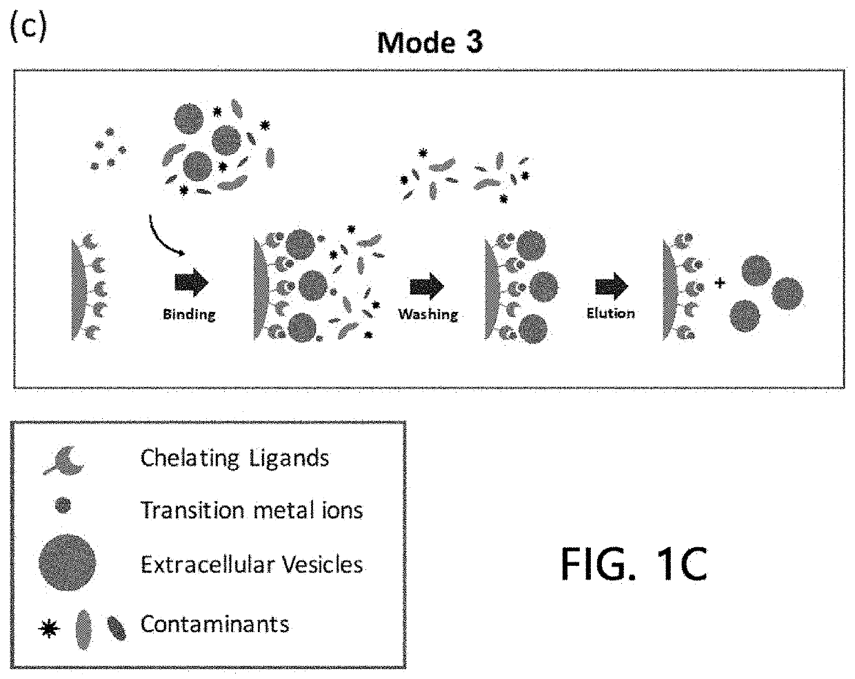
[0045]The extracellular vesicle isolation methods according to the present invention have advantages of being capable of efficiently isolating the extracellular vesicles while preserving the shape or properties thereof because costly equipment such as a centrifuge is not required and the samples are not exposed to an extreme environment during the isolation process. Further, the method of the present invention can be applied in combination with existing isolation methods, and can maximize the efficiency of extracellular vesicle isolation by applying the method of the present invention before or after performing the existing methods.
[0046]In addition, the extracellular vesicle isolation method of the present invention can rapidly and effectively isolate extracellular vesicles without any limitation in the amount of sample, and thus can play an important role in mass production of extracellular vesicles, while being utilized for clinical diagnosis by applying the method of the present invention for pre-treating and post-treating a small amount of body fluid samples.
[0047]Furthermore, the extracellular vesicle isolation method of the present invention can fractionate the extracellular vesicles into their subsets using various metals based on the fact that the affinity of the extracellular vesicles for a specific metal varies depending on the types of the extracellular vesicles. The fractionated extracellular vesicle subsets can be utilized for multidimensional disease diagnosis, and can solve the problems of the existing diagnostic markers and enable various applications by applying the previously developed various disease diagnostic markers to the method of the present invention.

Problems solved by technology

However, when ultra-centrifugation is used for isolating the extracellular vesicles, it presents such limitations as excessive amount of labor and time required due to its complex steps, the necessity of heavy equipment, and a large amount of a sample required for its tasks.
Thus, ultra-centrifugation is not suitable for a clinical diagnosis which requires a rapid result using a small amount of a sample and a therapeutic method in which a large amount of the extracellular vesicles is needed.
Size exclusion is usually used with ultra-centrifugation, and thus has an advantage in that the purity of extracellular vesicles may be increased, but has a limitation in that extracellular vesicles can be attached to a filter, leading to a low yield rate after isolation.
Immunoaffinity isolation is a method for isolating extracellular vesicles by attaching antibodies to the extracellular vesicles, and has an advantage of a significantly high specificity, while being problematic since it is not suitable for practical diagnosis and treatment due to extensive time and costs to produce antibodies.
However, a method for isolating extracellular vesicles using the affinity between metal ions and extracellular vesicles has not been reported yet.
The fundamental problem of these efforts lies in the isolation of extracellular vesicles from living tissues or body fluids, while it is almost impossible to purify the extracellular vesicles in a body fluid exhibiting a relatively limited amount and high complexity by conventional methods.

Method used


Image

  • Extracellular vesicle isolation method using metal
  • Extracellular vesicle isolation method using metal
  • Extracellular vesicle isolation method using metal

Examples


example 1

ion and Analysis of Sample Extracellular Vesicles

[0078]1-1. Isolation and Purification of Sample Extracellular Vesicles

[0079]Remaining cells and precipitate were removed by centrifugation (repeated twice in total) of a freshly obtained culture solution of a colorectal cancer cell line SW480 at 500×g for 10 minutes. A precipitate was removed again by centrifugation (repeated twice in total) of the supernatant at 2,000×g for 20 minutes.

[0080]In order to perform the Pt purification and precipitation of extracellular vesicles, which is present in the supernatant, a solution for inducing precipitation of extracellular vesicles (8.4% polyethylene glycol 6000, 250 mM NaCl, 20 mM HEPES, and pH 7.4) was added to the supernatant and the resulting mixture was stored in a refrigerator for 16 hours, the precipitated extracellular vesicles were then harvested by centrifugation at 12,000×g for 30 minutes and dissolved in a HEPES-buffered saline (20 mM HEPES, 150 mM NaCl, and pH 7.4).

[0081]In order...

example 2

spect of Sample Extracellular Vesicles to Stationary Phase in the Absence of Metal

[0092]After sample extracellular vesicles (4×10¹⁰ particles) were loaded onto a column (1 ml) filled with a stationary phase (IDA-Sepharose) using HEPES buffer solution for 5 minutes using an HPLC system, they were washed with the same buffer solution for another 5 minutes. Then, chromatography was attained on column with concentration gradient at a pH of 7.2 using 0 to 500 mM imidazole / HEPES buffer solution for a 20 column volume, and eluates of all the processes including binding and washing processes were fractionated into 1-ml fractions, respectively.

[0093]A binding aspect of extracellular vesicles to the stationary phase was analyzed by performing a chromatogram analysis (absorbances at 260 nm and 280 nm were measured, respectively), a nanoparticle analysis, and sandwich ELISA on the fractionated eluate by the methods as described above, respectively. The results are illustrated in FIGS. 7 and 8. ...

example 3

of Affinity of Sample Extracellular Vesicles for Cu(II)

[0094]After 0.5 ml of 500 mM Cu(II) ions were bound to a column (1 ml) filled with a stationary phase (IDA-Sepharose) using an HPLC system, non-bound residues were washed off with HEPES buffer solution of a 20 column volume.

[0095]After sample extracellular vesicles (4×10¹⁰ particles) were loaded onto the Cu(II) ions-bound stationary phase column using HEPES buffer solution for 5 minutes, the sample extracellular vesicles were washed with the same buffer solution for another 5 minutes. Eluates of all the processes including binding and washing processes were fractionated into 1-ml fractions, respectively, with a concentration gradient at a pH of 7.2 using 0 to 500 mM imidazole / HEPES buffer solution for a 20 column volume. A binding aspect of extracellular vesicles to the stationary phase was analyzed by performing a chromatogram analysis (absorbances at 260 nm and 280 nm were measured, respectively), a nanoparticle analysis, and ...


PUM

PropertyMeasurementUnit
sizesaaaaaaaaaa
pHaaaaaaaaaa
pHaaaaaaaaaa

Abstract

An extracellular vesicle isolation method using a metal, and a method for isolating extracellular vesicles from various samples using metal affinity are disclosed. An extracellular vesicle isolation method has the advantages of not requiring costly equipment, of being able to be applied without limits on sample quantity, and of being capable of efficiently isolating extracellular vesicles while preserving the shape or properties thereof. Moreover, the method can be combined with existing isolation methods to maximize the efficiency of extracellular vesicle isolation, and can be utilized in disease diagnosis, disease treatment, multi-omics research, and extracellular vesicle property research and the like using the isolated extracellular vesicles.

Description

TECHNICAL FIELD[0001]The present invention relates to a method for isolating extracellular vesicles by using a metal, and more particularly, relates to a method for isolating extracellular vesicles from various samples using a metal affinity.BACKGROUND ART[0002]Extracellular vesicles are nano-sized vesicles naturally secreted by all living organisms or cells from humans to bacteria through a universal cellular mechanism. Extracellular vesicles derived from eukaryotic cells have been reported to be involved in the differentiation of erythrocytes, the regulation of immune responses, and the like, and play an important role in the progression, metastasis, angiogenesis, and the like of cancer particularly in the micro environment of cancer cells. Thus, the extracellular vesicles have been drawing much attention in their applicability as a diagnostic marker for various diseases including cancer.[0003]It has been reported that extracellular vesicles secreted by prokaryotic cells contain t...

Claims


Application Information

Patent Timeline
Patent Type & Authority Patents(United States)
IPC IPC(8): B01D15/38B01D15/42B01J20/32B01D15/36B01D15/34C12N1/16C12N1/20C12N5/071
CPCB01D15/3828B01D15/424B01J20/3265C12N1/16C12N1/20C12N5/0602B01D15/34B01D15/361B01D15/3804C12N2509/00G01N33/54306C12N5/0696
Inventor GHO, YONG SONGLEE, CHANG JINPARK, HYUN TAEK
Owner ROSETTA EXOSOME
Why Eureka
Try Eureka