
Iron platinum particles for adherence of biologics on medical implants

a technology of iron platinum particles and biologics, applied in the field of localized cell attraction, capture and retention in vivo, to achieve the effect of avoiding magnetization in situ

Active Publication Date: 2022-03-29
YALE UNIV
View PDF100 Cites 0 Cited by
  • Summary
  • Abstract
  • Description
  • Claims
  • Application Information

AI Technical Summary

Benefits of technology

[0011]It has been discovered that iron / platinum (Fe / Pt) particles can be dispersed in a polymer and coated into or onto, or directly linked to, medical devices and magnetized. The magnetized devices are used to attract, capture, and / or retain magnetically labeled cells on the surface of the device in vivo. The magnetic devices are particularly useful for capturing and retaining cells exposed to the stresses and forces of biological fluid flow, for example, on stents implanted following angioplasty, to form a patent endothelial surface to avoid restenosis from occurring. They can also be used to increase tissue integration at the site of implantation of a prosthesis such as a metal surface hip or knee prosthetic, or decrease bone erosion surrounding a metal bone screw, pin or plate. The devices may be made of a metal, polymer or combination thereof.
[0021]Methods of treatment using the disclosed devices are also provided. For example, a method of treating a vascular injury can include implanting a magnetic stent into a subject in need thereof at or adjacent to the site of injury, and administering to the subject an effective amount of magnetic endothelial cells, endothelial precursor cells, or other pluripotent cells or stem cells to increase or enhance repair at the site of injury. A method of treating atherosclerosis can include implanting a magnetic stent into a subject in need thereof at a site of atherosclerosis. Such a method can also include administering to the subject an effective amount of magnetic endothelial cells or endothelial precursor cells to reduce or prevent restenosis at the site of stent deployment. Some embodiments further include co-administering the subject therapeutic, prophylactic or diagnostic agent(s) with the magnetic cells to enhance or increase repair of vascular injury, reduce or prevent restenosis, early or late thrombosis and / or reduce or prevent neointima formation.

Problems solved by technology

Further, there are advantages to avoiding magnetization in situ, since magnetization in situ may cause the device to move, which may cause physiological problems.

Method used


Image

  • Iron platinum particles for adherence of biologics on medical implants
  • Iron platinum particles for adherence of biologics on medical implants
  • Iron platinum particles for adherence of biologics on medical implants

Examples


example 1

ion of Magnetized Stents for Treating Restenosis

Materials and Methods

[0151]Synthesis of Iron / Platinum Particles

[0152]The synthesis involves simultaneous chemical reduction of Pt(acac)2 and Fe(acac)3 by 1,2-hexadecanediol at high temperature (250° C.) in solution phase. The synthesis was handled under standard airless techniques in an argon atmosphere. The reagents were obtained from commercial sources and used without further purification. A mixture of 0.5 mmol of Pt(acac)2, 1.0 mmol of Fe(acac)3, and 1,2-hexadecanediol (5.0 mmol) was added to a 125 mL European flask containing a PTFE coated magnetic stir bar. Dioctyl ether (30 mL) was then transferred into the flask and the contents stirred while purging with Ar for 20 min at room temperature. The flask was then heated to 100° C. and held at 100° C. for 20 min. During this hold, 0.05 mmol (0.17 mL) of oleylamine and 0.05 mmol (0.16 mL) of oleic acid were injected into the flask while continuing the Ar purge. After the 20 min hold, ...

example 2

d Cells are Retained on Magnetized Stents In Vitro

[0187]Materials and Methods

[0188]Dye release

[0189]Coating stability of polymer layer in the presence of Fe / Pt particles, iodinated dendrimer (ID) particles or both was studied by measuring dye release. Rhodamine B (1 wt. %) was dissolved in the PLA chloroform solution and the particles were added to the solution. The solutions with or without particles were dropped on the cover glasses and dried overnight. The cover glasses were incubated in PBS at 37° C. and PBS (1 mL) was taken to measure released dye at the desired time points.

[0190]Body Clearance

[0191]PLGA particles encapsulating DiR dye ((1,1′-Dioctadecyl-3,3,3′,3′-Tetramethylindotricarbocyanine Iodide), Life Technologies) and / or Fe / Pt were prepared with a typical emulsion method. Briefly, PLGA (75 mg), Fe / Pt (25 mg), and dir dye (1 mg) were dissolved in chloroform, and then added drop-wise to 5% polyvinyl alcohol (PVA). The mixture was sonicated three times and then added to 0....

example 3

tion of Magnetized Cells in PLGA Particles

[0200]Cells (such as endothelial cells, macrophages or progenitor cells) can be made magnetically susceptible by intracellular incorporation of iron oxide or attachment to the surface.

[0201]Materials and Methods

[0202]To facilitate enhanced loading of iron-oxide in cells. PLGA particles are fabricated by the double emulsion method encapsulating a high concentration of iron oxide and a dye (Coumarin 6). PLGA particles encapsulating hydrophobic superparamagnetic iron oxide (SPIO) were prepared and surface-functionalized with avidin-palmitic acid. Briefly, PLGA (107 mg) and hydrophobic SPIO (26 mg) were dissolved in chloroform (2 mL) and then added drop-wise to a vortexing solution of 5% PVA (4 mL) and the resulting mixture was sonicated three times for 10 s at an amplitude of 38% (400 W). The mixture was then added drop-wise to 100 mL of 0.2% PVA and left stirring for 3 h to evaporate the solvent. Particles were collected by centrifugation at 1...


PUM

PropertyMeasurementUnit
temperatureaaaaaaaaaa
thicknessaaaaaaaaaa
thicknessaaaaaaaaaa

Abstract

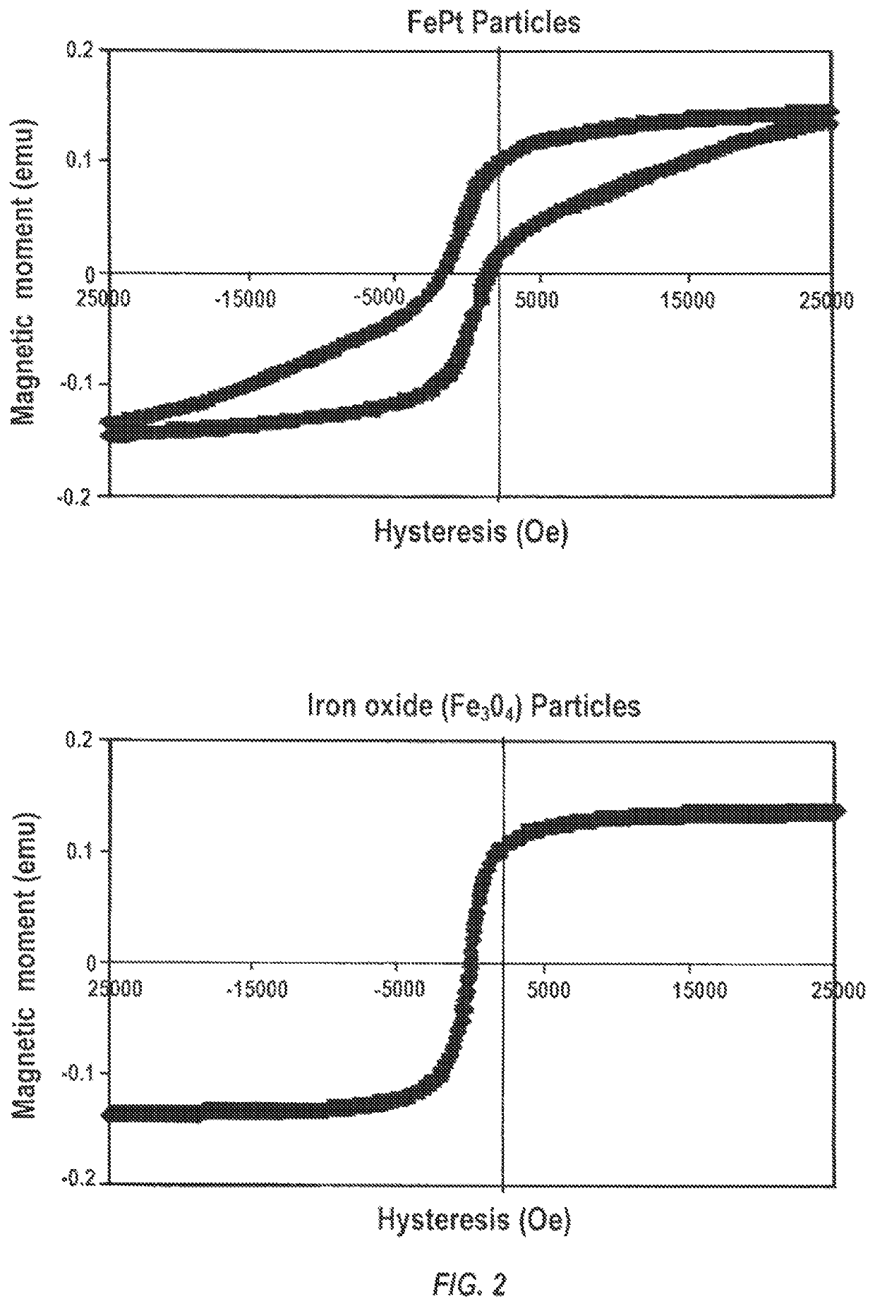
It has been discovered that iron-platinum ferromagnetic particles can be dispersed in a polymer and coated into or onto, or directly linked to or embedded on to, medical devices and magnetized. The magnetized devices are used to attract, capture, and / or retain magnetically labeled cells on the surface of the device in vivo. The magnetic particles have an iron / platinum core. Annealing the Fe / Pt particle is very important for introducing a L10 interior crystalline phase. The Fe:Pt molar ratio for creation of the crystal phase is important and a molar range of 1.2-3.0 Fe to Pt (molar precursors, i.e. starting compounds) is desired for magnetization. The magnetic force as a whole can be measured with a “Super Conducting Quantum Interference Device”, which is a sensitive magnetometer. The overall magnetic force is in the range from 0.1 to 2.0 Tesla.

Description

STATEMENT REGARDING FEDERALLY SPONSORED RESEARCH[0001]This invention was made with government support under T32 grant Award Number 5T32HL098069 awarded by the National Institutes of Health. The government has certain rights in the invention.CROSS REFERENCE TO RELATED APPLICATION[0002]This application is a 371 application of PCT / US2015 / 023880, filed Apr. 1, 2015, which is incorporated herein by reference in its entirety.FIELD OF THE INVENTION[0003]The field of the invention is generally related to localized cell attraction, capture, and retention in vivo, and more particularly to compositions and methods for attracting, capturing, and retaining target cells to a metal surface of a device or graft, noninvasive or invasive tracking of cell accumulation to the device or graft, and noninvasive or invasive evaluation of device status.BACKGROUND OF THE INVENTION[0004]Cardiovascular disease is the one of the biggest killers worldwide. The direct and indirect costs to the European Union alon...

Claims


Application Information

Patent Timeline
Patent Type & Authority Patents(United States)
IPC IPC(8): A61K41/00A61N2/00A61N2/06A61K35/44A61K35/545A61L27/02A61L27/04A61L27/06A61L27/18A61L27/30A61L27/34A61L27/58A61L31/02A61L31/06A61L31/08A61L31/10A61L31/14A61F2/82
CPCA61K41/0052A61K35/44A61K35/545A61L27/025A61L27/042A61L27/047A61L27/06A61L27/18A61L27/306A61L27/34A61L27/58A61L31/022A61L31/028A61L31/06A61L31/088A61L31/10A61L31/148A61N2/002A61N2/06A61F2/82A61F2210/009A61F2210/0076A61L2400/18A61L2420/02A61L2420/06A61L2420/08A61L2430/20A61P43/00A61P9/08A61P9/10A61P9/14
Inventor FAHMY, TAREKSINUSAS, ALBERTLEE, JUNG SEOKKIM, DONGINMATHUR, ANTHONYMARTIN, JOHN
Owner YALE UNIV
Learn More