
Analytical process for predicting the therapeutic effect of BH3 mimetics

a technology of bh3 mimetics and analytic process, which is applied in the field of analytic process for predicting the therapeutic effect of bh3 mimetics, can solve the problems of limited life span of leukemic blast cells in vitro from peripheral blood, bone marrow or lymph nodes, and achieve the effect of reducing the risk of leukemic blast cells in vitro

Active Publication Date: 2022-05-17
MEDIZINISCHE HOCHSCHULE HANNOVER
View PDF7 Cites 0 Cited by
  • Summary
  • Abstract
  • Description
  • Claims
  • Application Information

AI Technical Summary

Benefits of technology

[0015]A BH3 mimetic is a pharmaceutical compound that mimics the binding of the BH3 domain of Bcl-2 (B-cell lymphoma 2) family proteins. Pro-apoptotic BH3 only proteins such as BIM bind to the BH3 binding groove in pro-survival proteins such as Bcl-2, Bcl-XL or Mcl-1. Accordingly, BH3 mimetics bind to and block the BH3 binding groove within pro-survival proteins thereby facilitating induction of mitochondrial apoptosis.
[0018]The analytical process can be carried out within a short period of time so that the short in vitro survival time of leukemic cells is not hampering the analysis. Further, the analytical process avoids the analysis of cellular proteins or of genetic aberrations. Therefore, the analytical process is useable for direct prediction of the clinical response of an individual patient to a specific BH3 mimetic without need for analysis of surrogate markers such as e.g. molecular analyses of tumour markers or cytogenetics and molecular genetics as used in e.g. certain solid tumours.
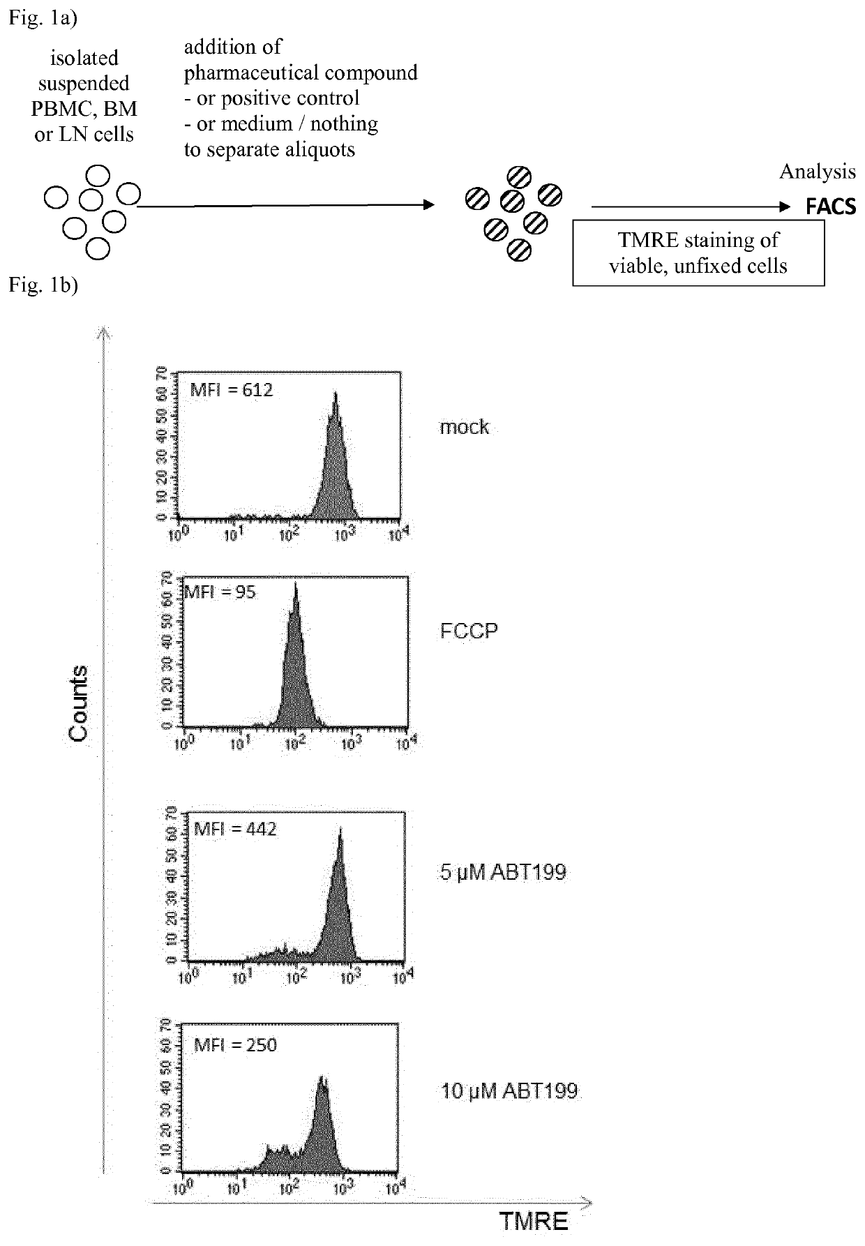
[0024]Optionally additionally, the fluorescence of the dye for measuring transmembrane potential of mitochondria is determined for CD19− (CD19 negative) cells, e.g. in order to determine the proportion of live non-tumour cells, e.g. on CD19− cells, that undergo MOMP caused by the pharmaceutical compound, e.g. for assessment of unwanted side effects of the pharmaceutical compound. This allows quantification of the therapeutic window and adverse effects of the pharmaceutical compound on normal cells, e.g. non-tumour cells, in one FACS analysis. Generally, the process includes at least one aliquot of the mononuclear cells treated in parallel with no pharmaceutical compound added as a negative control, and preferably at least one aliquot of the mononuclear cells treated in parallel with a compound known to induce MOMP, preferably known to induce MOMP independent from BH3-mimetics and within a short time as a positive control, e.g. FCCP.
[0028]More preferably, subsequent to the process being carried out on an initial sample, the process is additionally carried out on at least one sample, which originates from the same patient at a later point in time, e.g. for monitoring the efficacy of at least two different pharmaceutical compounds, which preferably are BH3 mimetics, and the determination of the pharmaceutical compound which has the more intense efficacy against the leukemia or lymphoma cells of the sample originating from the same patient at a later point in time. In this embodiment, the process allows to monitor the efficacy of the BH3 mimetics and to select a pharmaceutical compound, which preferably is a BH3 mimetic, having a more intense efficacy also e.g. in samples originating from a patient who shows a decline in the efficacy of the pharmaceutical compound, e.g. BH3 mimetic, used for treatment, e.g. in samples originating from patients showing resistance against treatment with a certain compound, preferably a BH3 mimetic, e.g. samples from refractory patients. This allows monitoring and selection of effective pharmaceutical compounds, especially of BH3 mimetics, during the course of therapy for individual patients.
[0032]The process has the advantage of a short duration, so that the desired result of the effectiveness of the pharmaceutical compound added to the isolated PBMC, BM or LN mononuclear cells is obtained within a short period of time, e.g. in under 4 h, preferably in under 3 h, more preferably in under 2 h or in under 1 h, starting from isolated PBMC, BM or LN mononuclear cells and does not interfere with the short survival time of leukemic blast cells in vitro. This short duration of the process is advantageous, especially for acute forms of leukemia, as it allows the administration of the pharmaceutical compound to the patient after the short duration, without a longer delay. The blood, BM or LN sample originates from a patient who was diagnosed as or is suspected of having leukemia, especially chronic lymphatic leukemia (CLL), preferably acute myeloic leukemia (AML) or acute lymphatic leukemia (ALL) and advanced chronic myeloid leukemia (CML) or all kind of lymphoma. The blood, BM or LN sample originating from a patient generally can be drawn prior to the analytical process.
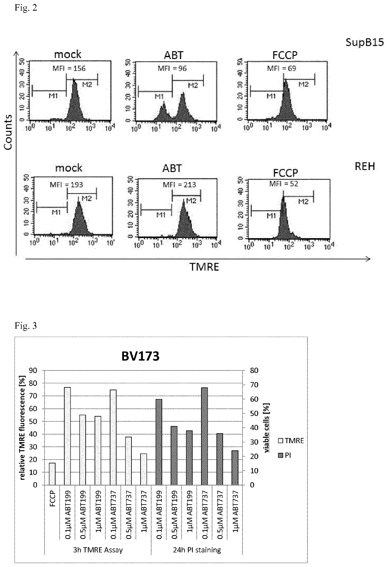
[0034]It has been found that the analytical process determines the effectiveness of a pharmaceutical compound in a concentration-dependent manner, which is interpreted as an indication that the process only determines the effectiveness in respect of inducing apoptosis in isolated PBMC, BM or LN mononuclear cells without additional effects caused by a higher concentration of the pharmaceutical compound. Therefore, the analytical process can be done at concentrations of the pharmaceutical compound higher than the pharmacological concentrations adjusted in the patient. For example, the concentration of the pharmaceutical compound in the process can be higher by a factor of at least 2 or a factor of at least 5 or by a factor of at least 10, preferably higher by a factor of at least 100, more preferred higher by a factor of at least 1000 than the pharmacological concentrations adjusted in the patient. The pharmacological concentrations adjusted in the patient can e.g. be determined by the recommended dosage of the pharmaceutical compound. At higher concentrations of the pharmaceutical compound, the process can be performed in a shorter duration, e.g. in less than 1 h, e.g. within 40 min, preferably within 30 min, starting from the isolated PBMC, BM or LN cells, i.e. measured from the end of the isolation of the respective cells. Furthermore, the induction of MOMP in isolated leukemic PBMC, BM or LN mononuclear cells by the compound, which preferably is a BH3 mimetic, always precedes and leads to cellular apoptosis under physiologic conditions so MOMP induction can be used as a reliable predictor for cytotoxicity of the compound analysed.

Problems solved by technology

It is known that leukemic blast cells from the peripheral blood, bone marrow or lymph nodes in vitro have a very limited life span, in particular ALL-blasts e.g. after isolation.

Method used


Image

  • Analytical process for predicting the therapeutic effect of BH3 mimetics
  • Analytical process for predicting the therapeutic effect of BH3 mimetics
  • Analytical process for predicting the therapeutic effect of BH3 mimetics

Examples


example 1

NT OF SUSCEPTIBILITY OF CELL LINES FOR APOPTOSIS

[0053]As representatives for isolated PBMC, BM or LN mononuclear cells, the B-ALL cell lines SUP-B15, which expresses BCR-ABL (BCR-ABL-positive), and the BCR-ABL negative REH were used in the analytical process in suspension in cell culture medium RPMI1640. As a positive control for MOMP, FCCP was added to 5 μM to one aliquot, as a negative control an aliquot of the suspended cells was used with addition of the same volume of physiological saline, and ABT-199 was added to 10 nM final concentration. After incubation at 37° C. for 3 h, TMRE was added to a final concentration of 50 nM and the aliquots were incubated for further 15 min at 37° C., in a 5% CO2 atmosphere each time. The measurement results by FACS are shown in FIG. 2. For the aliquots containing ABT-199, the MFI is reduced by approx. 40% in the BCR-ABL positive SUP-B15 cells, but for the BCR-ABL negative REH cells, the MFI essentially remains unaffected by ABT-199 in comparis...

example 2

NT OF SUSCEPTIBILITY OF PBMC FOR APOPTOSIS

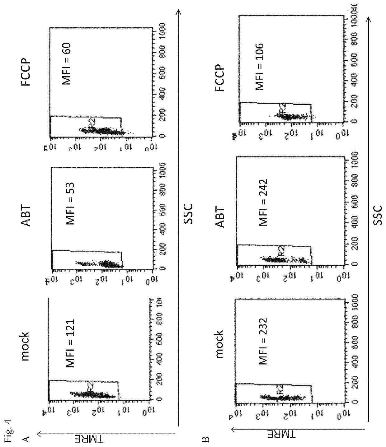
[0055]PBMC were isolated by density gradient centrifugation using Biocoll from the heparinized blood sample of a patient diagnosed to have CLL (ABT-199, Venetoclax, is approved for treatment of CLL with 17p-deletion). As a comparison, PBMC from a healthy donor were isolated. PBMC were suspended in RPMI1640 cell culture medium and transferred as aliquots into separate tubes, to which ABT-199 was added to a final concentration of 1 μM and incubated at 37° C. for 3 h in a 5% CO2 atmosphere. Subsequently, TMRE was added to 50 nM and aliquots were further incubated for 30 min, then analysed by FACS according to the invention. As a negative control (mock), physiological saline was added, as a positive control FCCP to 5 μM (FCCP).

[0056]FIG. 4A shows the results for PBMC from a CLL patient, FIG. 4B shows the results for the PBMC from healthy blood sample. The results demonstrate that after 3 h incubation, the PBMC from the CLL blood sample, the pres...

example 3

NT OF SUSCEPTIBILITY OF PBMC FOR APOPTOSIS

[0058]PBMC or BM mononuclear cells were isolated by density gradient centrifugation from blood or bone marrow samples originating from patients who were newly diagnosed as ALL (n=12), from blood or bone marrow samples originating from CML patients in chronic phase (CML-CP, n=7), and from blood samples obtained from healthy volunteers (n=6). In accordance with the analytical process, PBMC or BM cells were incubated in cell culture medium with ABT-199 added to 1 μM for 3 h at 37° C. in a 5% CO2 atmosphere for cell culture conditions, followed by addition of TMRE to 50 nM and incubation for another 30 min, then analysed by FACS for fluorescence by TMRE.

[0059]The results are shown in FIG. 5, indicating essentially no induction of apoptosis for the PBMC from healthy donors, a strong induction of apoptosis for the PBMC or BM cells from these ALL samples (ALL), and essentially no induction of apoptosis for the PBMC or BM cells from the CML-CP patie...


PUM

PropertyMeasurementUnit
survival timeaaaaaaaaaa

Abstract

The present invention relates to an analytical in vitro process for predicting the therapeutic effectiveness of at least one pharmaceutical compound in the treatment of leukemia and / or lymphoma, the process analysing the transmembrane potential of mitochondria in cells isolated from a patient by quantification of fluorescence emitted from a dye indicating induction of apoptosis.

Description

[0001]The present invention relates to an analytical process for predicting the therapeutic effectiveness of a pharmaceutical compound, which preferably is a BH3 mimetic, in the treatment of leukemia, including chronic lymphatic leukemia (CLL), preferably in acute myeloic leukemia (AML) or in acute lymphoblastic leukemia (ALL) as well as advanced chronic myeloid leukemia (CML) or lymphoma, including diffuse large B-cell lymphoma (DLBCL), Burkitt-lymphma, follicular lymphoma, mantle cell lymphoma, splenic marginal zone lymphoma, lymphoblasmocytic lymphoma, nodal marginal zone B-cell lymphoma, mediastinal large B-cell lymphoma, plasma cell myeloma, angioimmunoblastic T-cell lymphoma, anaplastic large cell lymphoma, T-cell prolymphocytic leukemia, T-cell LGL leukaemia, aggressive NK cell leukaemia, adult T-cell leukaemia / lymphoma, extranodal NK / T cell lymphoma (nasal type), enteropathy-type T-cell lymphoma, hepatosplenic T-cell lymphoma, subcutaneous panniculitis-like T-cell lymphoma, ...

Claims


Application Information

Patent Timeline
Patent Type & Authority Patents(United States)
IPC IPC(8): C12Q1/00G01N33/50
CPCG01N33/5011G01N33/57407G01N33/57426G01N2800/52
Inventor SCHERR, MICHAELAEDER, MATTHIAS
Owner MEDIZINISCHE HOCHSCHULE HANNOVER
Learn More