Medical imaging apparatus and method for the imaging of a light-sensitive object, such as biological tissue

a technology of medical imaging and light-sensitive objects, applied in the field of medical imaging apparatus and method for the imaging of light-sensitive objects, can solve the problems of discomfort for patients, complicated observation and imaging of objects such as living tissue, and even damage to tissue by illumination

Active Publication Date: 2022-11-15
LEICA INSTR SINGAPORE PTE
View PDF34 Cites 0 Cited by
  • Summary
  • Abstract
  • Description
  • Claims
  • Application Information

AI Technical Summary

Benefits of technology

[0011]The method and apparatus according to the invention have the advantage of lessened discomfort for the patient and significantly reducing the risk of injury to the tissue. The inventive method and apparatus allow for significantly longer examinations, which helps to prevent errors in diagnosis and therapy.
[0015]The input images and the output images may be part of a time-series of input images and output images which are captured and / or displayed at a frame rate which is higher than the flicker fusion rate so that any change in the tissue during observation can be smoothly rendered in the output images.
[0019]According to another advantageous embodiment of the invention, the medical imaging apparatus may comprise an illumination system for illuminating the object with light including an illumination set of spectral bands. The illumination spectral bands may be bands at least partly in the non-visible light range, in particular in at least the input spectral bands. The output-only spectral band preferably is attenuated or not present in the illumination set. The illumination of the tissue in the input spectral bands ensures a good quality of the input images with respect to depth of field and spatial resolution without the need for special low-light optics or cameras. In the input spectral bands, there is no risk of injuring the tissue or putting the patient in discomfort. Further, in ophthalmological examinations, the eye will not react to an illumination in the non-visible light range by contracting the iris thus allowing for an easier inspection.
[0025]The camera may be an imaging spectroscope, in particular a multispectral camera or a hyperspectral camera. The more spectral bands used, the better the spectral resolution of the input spectral signature and the more accurate the assignment of the spectral signature of the object to the output set will be. For some applications a standard color camera e.g. operating the RGB color space may be sufficient.

Problems solved by technology

The observation and imaging of objects such as living tissue is complicated if the object is light-sensitive.
In some instances, the illumination may even damage the tissue.
In ophthalmology, the sensitivity of the light-sensitive tissue in the eye results in discomfort for the patient, a higher risk of injury and additional difficulties due to the eye's reflex reaction to light.
Such cameras are, however, expensive and bulky and need expensive and large high-quality optics.
The large aperture of the optics, which is needed to capture as much of the incoming light as possible, leads only to a limited depth of the field of view.

Method used


Image

  • Medical imaging apparatus and method for the imaging of a light-sensitive object, such as biological tissue
  • Medical imaging apparatus and method for the imaging of a light-sensitive object, such as biological tissue

Examples


Embodiment Construction

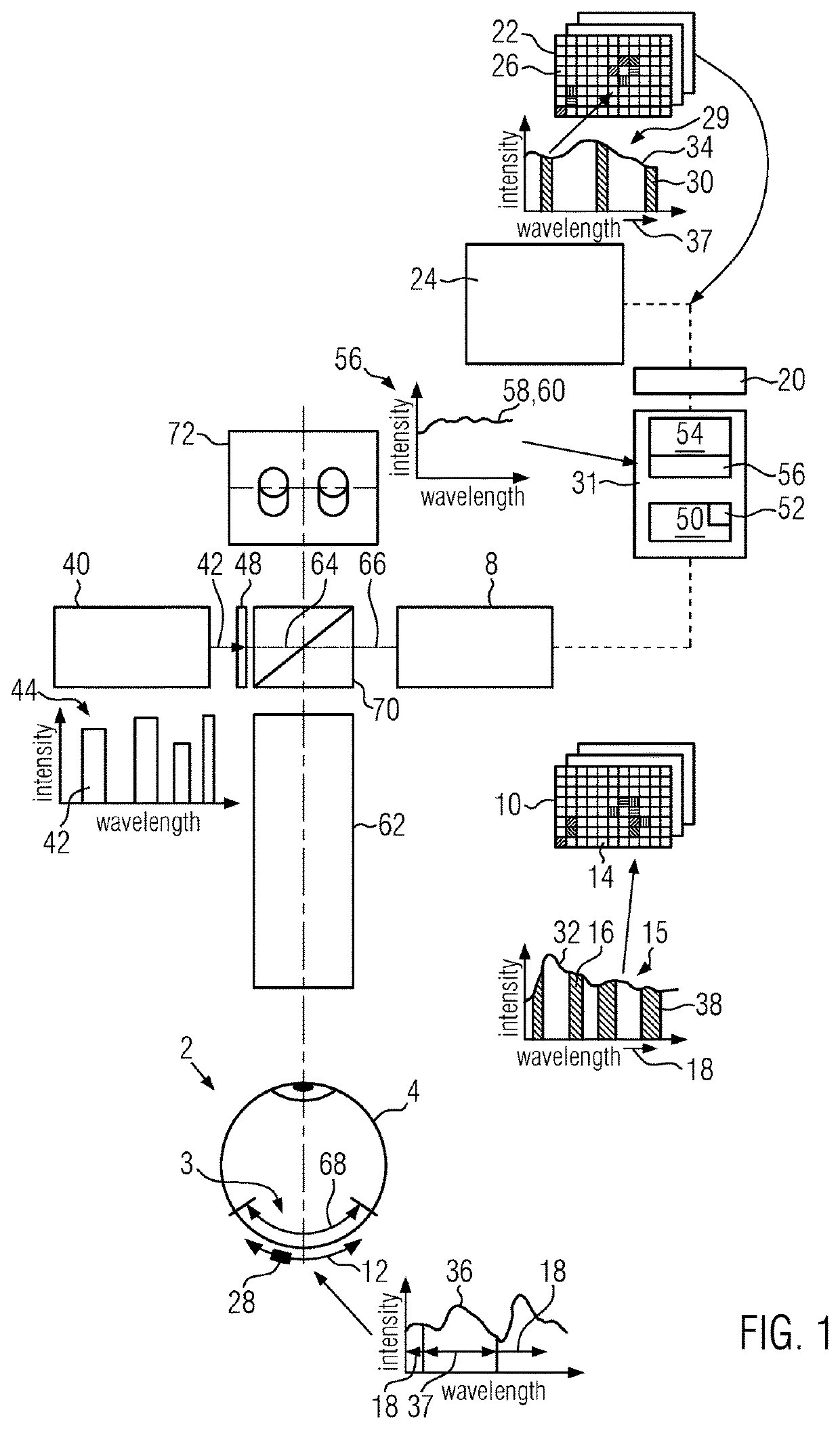
[0045]First, an example of a medical imaging apparatus 1 is described with reference to FIG. 1.

[0046]The medical imaging apparatus 1 is used for the imaging of an object 2, such as biological tissue 3, in particular light-sensitive regions of tissue, such as in an eye 4. The medical imaging apparatus 1 is in particular used in ophthalmology as an ophthalmological imaging device 6.

[0047]The medical imaging apparatus 1 comprises a camera 8 for capturing input images 10 of the tissue 3 located in a field of view 12. The camera 8 may be a color camera such as an RGB camera or an imaging spectrograph such as a multispectral or a hyperspectral camera.

[0048]Each input image 10 comprises input pixels 14. Each pixel 14 represents a region of the field of view. Each input image 10 and each input pixel 14, respectively, contains an input set 15 of at least spectral bands 16.

[0049]The camera 8 is sensitive in the at least two discrete input spectral bands 16 in the non-visible light-range 18. T...


PUM


Abstract

A medical imaging apparatus (1) and method for imaging a light-sensitive object (2), such as tissue (3) of the eye (4), displays the object (2) in spectral bands (30) of the visible light range (37) in which the object is sensitive and cannot be illuminated. The apparatus (1) has a camera (8) for capturing input images (10) in which a spectral signature (36) of the object is represented by an input set (15) of spectral bands (16). The apparatus generates output images (22) in which the spectral signature (60) in the visible light range (37) is represented by an output set (29) of spectral bands (30). Replacing at least one input-only spectral band (38) in the input set (15) by at least one output-only spectral band (39) in the output set (29) enables capture of input images (10) in spectral bands (16) where the object (2) has less sensitivity, such as in the non-visible light-range (18), and display of the object (2) in spectral bands (30) where the object would be too sensitive for illumination (40).

Description

CROSS REFERENCE TO RELATED APPLICATIONS[0001]This application claims priority of European patent application number 16181848.9 filed Jul. 29, 2016, the entire disclosure of which is incorporated by reference herein.FIELD OF THE INVENTION[0002]The invention relates to a medical imaging apparatus and method for the imaging of a light-sensitive object having a spectral signature, for example biological tissue, in particular light-sensitive tissue of a living body, such as of an eye. The medical imaging apparatus and method in particular are an ophthalmologic apparatus and method, respectively.BACKGROUND OF THE INVENTION[0003]The observation and imaging of objects such as living tissue is complicated if the object is light-sensitive. Light-sensitivity may cause the tissue to react if illuminated. This reaction may cover characteristics of the tissue that need to be observed to make a diagnosis. In some instances, the illumination may even damage the tissue.[0004]In ophthalmology, the se...

Claims


Application Information

Patent Timeline
Patent Type & Authority Patents(United States)
IPC IPC(8): G06K9/00G06T7/00A61B3/14A61B3/12A61B3/00
CPCG06T7/0012A61B3/0025A61B3/0041A61B3/12A61B3/14G06T2207/30041A61B3/0008A61B5/0075
Inventor THEMELIS, GEORGE
Owner LEICA INSTR SINGAPORE PTE
Why Eureka
Try Eureka