Method of detecting ultrasound contrast agent in soft tissue, and quantitating blood perfusion through regions of tissue

a contrast agent and soft tissue technology, applied in the field of detecting an ultrasound contrast agent, can solve the problems of reducing the detection accuracy of low concentrations of agents, affecting the detection accuracy of ultrasound contrast agents,

Inactive Publication Date: 2001-12-27
ANGELSEN BJORN
View PDF0 Cites 54 Cited by
  • Summary
  • Abstract
  • Description
  • Claims
  • Application Information

AI Technical Summary

Problems solved by technology

However, with the present wideband transducer technology it is only possible to utilize the second harmonic component of the signal by transmitting an ultrasound pulse with frequency spectrum in the lower part of the active frequency band of a wideband transducer.
Forward propagation of the ultrasound pulse through the tissue produces a distortion of the pulse due to pressure dependent propagation velocity.
The back scattered tissue signal then represents a background noise for the detection of the contrast agent, and hence limits the detectability of low concentrations of the agent based on the 2nd harmonic component of the back scattered signal.
Accordingly, considerable distortion of the scattered pulse directly in the scattering process results with high amplitudes in the 3rd and 4th harmonic component of the incident pulse frequency band.
There are however several practical problems in utilizing the 3rd and 4th harmonic component of the transmitted frequency band for detection and imaging of ultrasound contrast agent, as well as using such imaging to grade the degree of blood perfusion through tissue:
The first problem is that the present medical ultrasound transducers have so narrow a bandwidth that it is not possible to transmit a pulse with frequency band around f.sub.0, and receive back-scattered frequency components in the freq...

Method used


Image

  • Method of detecting ultrasound contrast agent in soft tissue, and quantitating blood perfusion through regions of tissue
  • Method of detecting ultrasound contrast agent in soft tissue, and quantitating blood perfusion through regions of tissue
  • Method of detecting ultrasound contrast agent in soft tissue, and quantitating blood perfusion through regions of tissue

Examples


Embodiment Construction

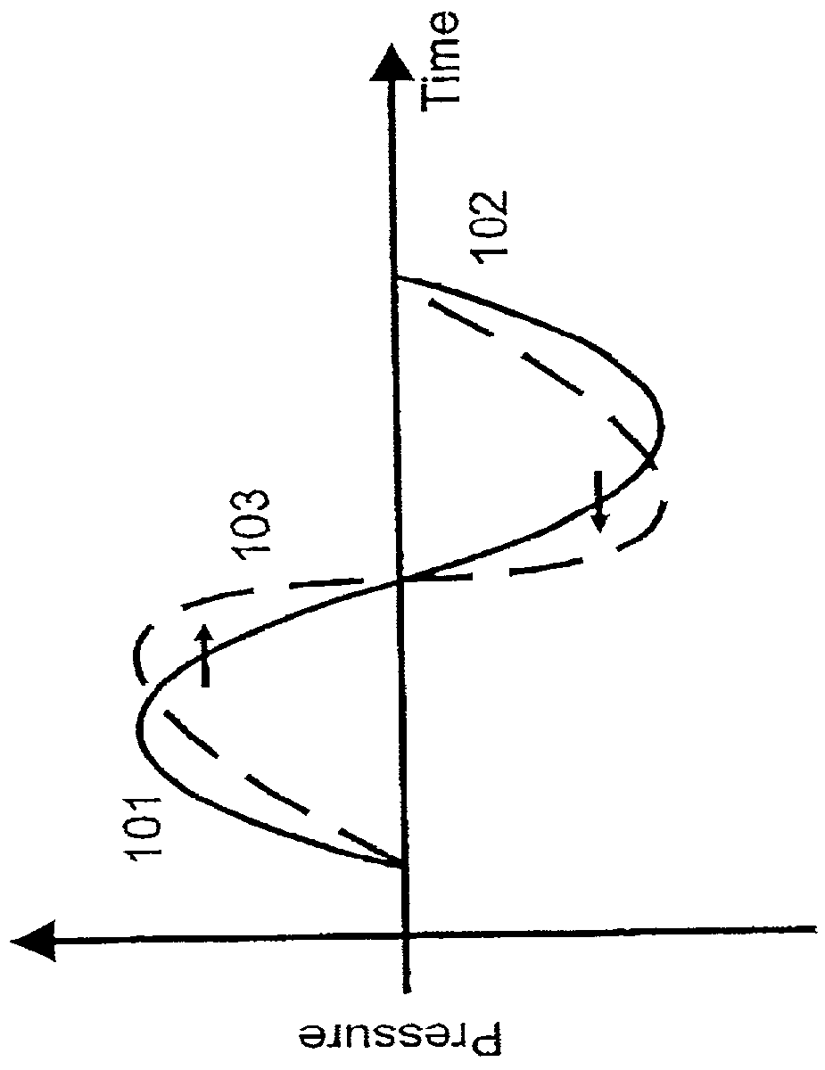
[0057] The stiffness of a soft tissue increases when the tissue is compressed by the positive pressure in a transmitted ultrasound pulse. The propagation velocity of an ultrasound wave therefore increases with positive pressure due to this stiffness increase. Similarly, the propagation velocity decreases with negative pressure in the incident pulse. The positive pressure swing 101 of the initial transmitted ultrasound pressure pulse in FIG. 1a hence propagates faster than the negative swing 102. After a propagation distance, we therefore get a distortion of the pressure pulse, for example illustrated as the dotted curve 103. The distortion of the pulse increases with the propagation distance.

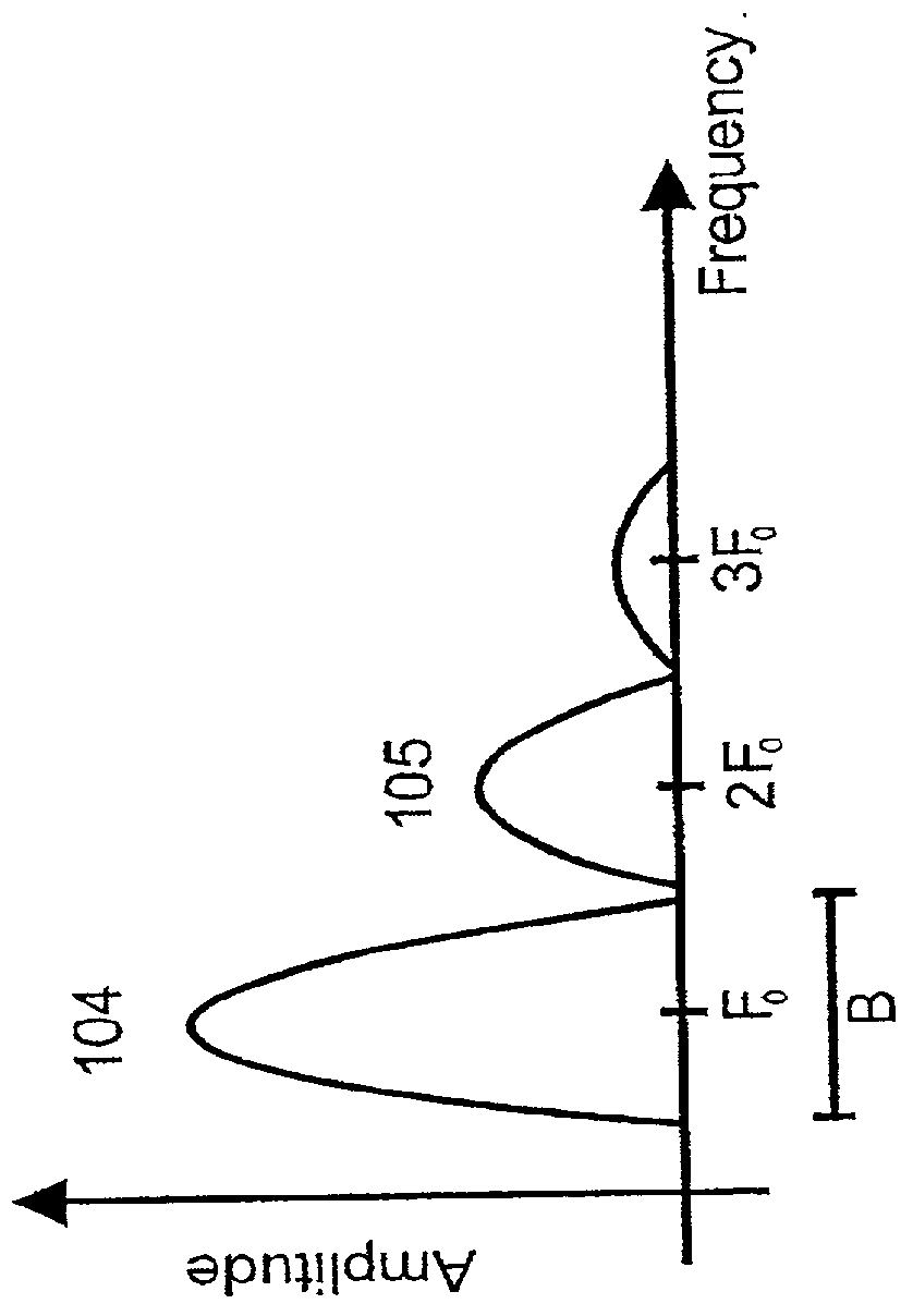
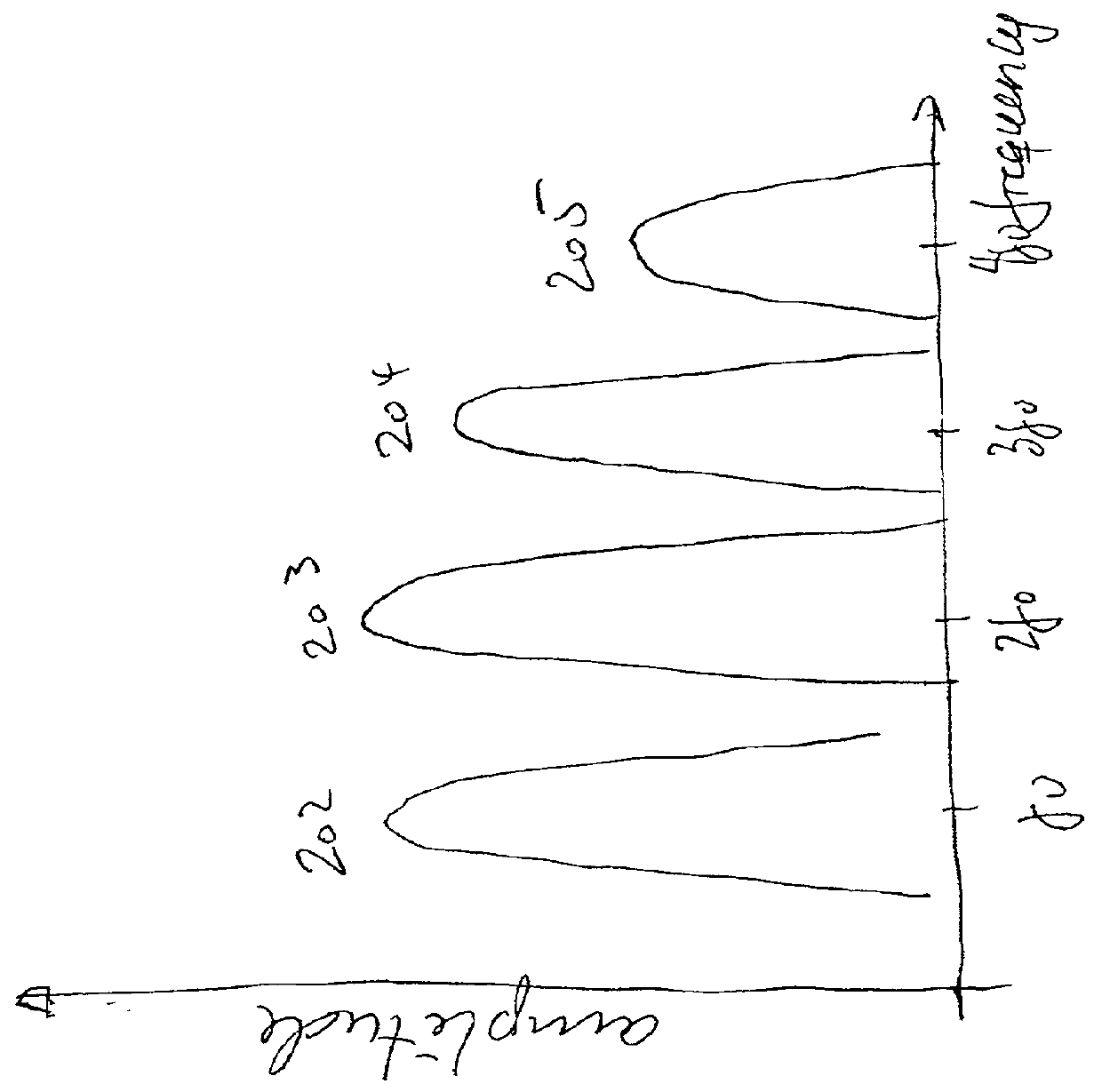
[0058] With no acoustic power absorption, the peak of the wave will eventually catch up with the trough, producing what is termed acoustic shock. However, the pulse distortion introduces higher harmonic frequency components in the pulse, with much larger absorption attenuation than the fundament...


PUM


Abstract

A method for detection of ultrasound contrast agent in soft tissue according to the present invention includes utilizing an ultrasound transmit beam former and transducer array assembly for transmitting directive, focussed ultrasound pressure pulses with steerable transmit amplitude, transmit aperture, transmit focus, and transmit direction, and with temporal frequency components within a limited band B centered at a frequency f0, towards a region of soft tissue that contains ultrasound contrast agent bubbles. The transmit pulse parameters are arranged, preferably using multiple transmit pulses, so that the incident pressure pulse that is utilized for imaging of the contrast agent for a particular depth, has minimal variation over the actual image range. The non-linearly distorted, back-scattered ultrasound signal is received from both the tissue and the ultrasound contrast agent bubbles with the same ultrasound transducer assembly and the received array element signals are passed through a receiver beamformer that has a steerable spatially directive receiver sensitivity.

Description

[0001] 1. Field of the Invention[0002] The present invention is directed to a method for detecting an ultrasound contrast agent in a soft tissue and quantitating blood perfusion through regions of tissue by detecting the contrast agent in the tissue.[0003] 2. Description of the Related Art[0004] An ultrasound contrast agent is a solution of small gas bubbles (diameter .about.5 .mu.m) that is injected into the blood stream. Such bubbles show strong and non-linear scattering of ultrasound at the frequencies used for medical ultrasound imaging. Medical applications of the contrast agents include, but are not limited to, enhancing imaging of blood vessels, improving the detection of the endocardium as a border of the ventricular cavities, and improving the detection of blood jets through leaking cardiac valves or septal defects.[0005] There has also been great hopes that ultrasound contrast agent should be able to detect and quantify blood perfusion through the tissue, especially the my...

Claims


Application Information

Patent Timeline
IPC IPC(8): A61B8/00A61B8/06G01S7/52
CPCA61B8/06A61B8/481G01S7/52038G01S7/52039
Inventor ANGELSEN, BJORN
Owner ANGELSEN BJORN
Why Eureka
Try Eureka