Fluid delivery and extraction device and method

a technology of extraction device and fluid, which is applied in the field of medical devices, can solve the problems of complex and perhaps life-threatening surgeries, trauma and tissue damage to the patient, and the restlessness of angioplasty patients

Inactive Publication Date: 2002-04-25
SUTURA
View PDF0 Cites 46 Cited by
  • Summary
  • Abstract
  • Description
  • Claims
  • Application Information

AI Technical Summary

Problems solved by technology

Invasive medical procedures typically cause trauma and tissue damage to the patient.
Moreover, these types of catheters have permitted clinicians to treat disorders with minimally invasive procedures that, in the past, would have required complex and perhaps life-threatening surgeries.
Unfortunately, roughly 30% of angioplasty patients experience restenosis (i.e., re-narrowing of a treated artery) following an angioplasty procedure.
If not diagnosed and treated quickly enough, restenosis can lead to stroke or death.
Conventional fluid delivery devices, however, fail to provide a means by which fluid compounds can be remotely injected directly into body tissue, such as the walls of blood vessels or fallopian tubes.

Method used


Image

  • Fluid delivery and extraction device and method
  • Fluid delivery and extraction device and method
  • Fluid delivery and extraction device and method

Examples


Embodiment Construction

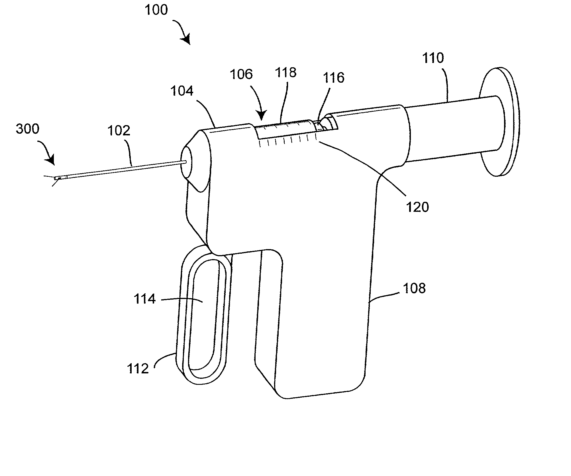
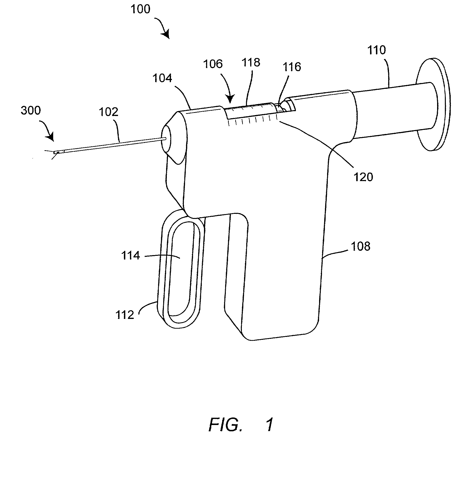
[0032] As shown in FIG. 1, an embodiment of a fluid delivery and extraction device 100 comprises a distal portion 300, a shaft 102, a main body 104, a window slot 106, a syringe 116, a luer connector 118, a plunger 110, a handle 108, a trigger actuator 112, a finger aperture 114, and a calibrated index 120. The shaft 102 is preferably flexible to allow it to bend when advanced through internal biological structures, particularly body lumens. The length of the shaft 102 may be modified to accommodate various fluid delivery and extraction applications. The trigger actuator 112 and the luer connector 118 are operatively connected to the distal portion 300 and may be used to remotely manipulate the components of the distal portion 300. The plunger 110 is operatively connected to the syringe 116 and may be used to remotely deliver or extract fluid through the distal portion 300. The window slot 106 enables the volume of fluid transferred to or from the syringe 116 to be visually monitore...


PUM


Abstract

A fluid delivery and extraction device enables the remote transfer of fluid to or from biological tissue. The device comprises an elongated member, a syringe movable relative to the elongated member and a pair of hypodermic needles movable relative to the elongated member. The elongated member has a distal portion which is adapted to be inserted into a biological structure. A steer wire or guide wire can be used to navigate the distal portion within cavernous biological structures, particularly body lumens. The distal portion has at least one retractable hypodermic needle that is configured to pierce the interior surface of a tubular biological structure and transfer fluid to or from the walls of the tubular biological structure. A physician can use a handle or other control mechanism provided at a proximal portion of the device to remotely move the hypodermic needles. A plunger can be used to transfer fluid through the hypodermic needles to or from the syringe. In operation, the hypodermic needles are deployed simultaneously or individually from the distal portion of the device. As the hypodermic needles move, they pierce the interior surface of the tubular biological structure. The hypodermic needles can also be configured to pass beyond the exterior surface of the tubular biological structure. The plunger is then used to transfer fluid either from the syringe to the patient or from the patient to the syringe. The hypodermic needles are then moved back to their retracted positions within the distal portion, and the device is withdrawn from the patient's body.

Description

[0001] This application claims the benefit of U.S. Provisional Application Serial No. 60 / 237,662, filed on Oct. 3, 2000, the entirety of which is hereby incorporated by reference.[0002] 1. Field of the Invention[0003] The present invention relates generally to medical devices. Specifically, the present invention relates to a fluid delivery and extraction device and method for transferring fluid to or from biological tissue that may not be directly accessible.[0004] 2. Description of the Related Art[0005] Invasive medical procedures typically cause trauma and tissue damage to the patient. Less invasive processes are ordinarily preferred, particularly when a biological structure to be medically treated is a body lumen, such as a blood vessel or a fallopian tube.[0006] Medical catheters such as balloon catheters have been proven efficacious in treating a wide variety of blood vessel disorders. Moreover, these types of catheters have permitted clinicians to treat disorders with minimall...

Claims


Application Information

Patent Timeline
Patent Type & Authority Applications(United States)
IPC IPC(8): A61B17/22A61B17/34A61M5/158A61M5/32
CPCA61B17/22A61B17/3478A61M2025/0086A61B2017/3445A61B2017/22077
Inventor NOBLES, ANTHONY A.ESKURI, ALAN
Owner SUTURA
Why Eureka
Try Eureka