Orthotic trauma device

a technology of orthotic device and pelvis, which is applied in the field of orthotic device, can solve the problems of inability to attempt to immobilize pelvic fractures by such means, inconvenient use, and additional harm, and achieve the effects of stabilizing the pelvis, reducing the movement of bone fragments, and reducing the volume of bled blood

Inactive Publication Date: 2002-10-17
PYNG MEDICAL CORP
View PDF0 Cites 100 Cited by
  • Summary
  • Abstract
  • Description
  • Claims
  • Application Information

AI Technical Summary

Benefits of technology

0010] The orthotic trauma device is suitable for use in the emergent treatment of most types of bone fractures and preferably pelvic fractures. Compression of the pelvis with this orthotic device stabilizes the broken bones of the pelvis and helps control blood loss by reducing the volume that can be bled into. Reducing the motion of the bone fragments allows bleeding sites to clot off helping to limit blood loss as well as greatly improving the comfort of the patient. Use of this device lessens the likelihood of further injury to nerves, blood vessels or internal organs when the patient is moved. These devices can easily be applied at the accident scene by emergency medical technicians or in an emergency room. These devices provide sufficient provisional stabilization of the pelvis that the need for emergent application of a pelvic external fixator is eliminated in most cases.
0011] This device does not interfere with the systematic evaluation of the trauma patient in the emergency room. It is preferably radiolucent and allows X-rays, CT scans, and angiography, abdominal ultrasound, peritoneal lavage, etc. to be carried out with the device in place. If need be it can be removed and reapplied easily. It can be left in place in most cases until workup is compete and definitive open reduction and internal fixation of the pelvic fracture can be performed.
0012] The orthotic devices of the present invention, include an orthosis body or bracing portion, in the shape of a wide belt, adapted to be wrapped around a portion of a body of a user, the bracing portion being elongated and formed from a material which is easily cut with cloth-cutting scissors and is also, preferably, formed from a material which is radiolucent (i.e., non-radiopaque). A detachable fastening device is provided at the distal or outer ends of the elongated bracing portion to detachably secure the ends around the part of the body where the bone fracture is located. The fastening device is preferably provided with a means for adjusting the tightness or tension of the bracing portion, accomplished preferably by increasing the mechanical advantage of the fastening device. Preferably the means for increasing the mechanical advantage of the fastening device is a detachable pulley system which includes a pair of pulley banks. A first bank of the pulley system is detachably disposed on a first distal end of the elongated bracing portion and a second pulley bank is detachably disposed on a second distal end of the elongated bracing portion. A cable is operatively connected to the two pulley banks such that the first and second banks of the pulley system are in juxtaposed relationship. The cable runs through a pulley on each juxtaposed pulley bank in series and in alteration, shortening of the cable pulling the two banks of pulleys and concomitantly the opposed ends of the orthosis body together and tightening the orthotic device with the aid of mechanical advantage dependent upon the number of pulleys mounted in the pulley bank on each opposing orthosis body end.
0013] In a preferred embodiment, each set of pulleys comprises two modular banks of pulleys which are detachably secured to opposing free ends of the same bracing portion. A cable is provided to connect pulleys in the opposing banks of pulleys in a set in series and in alteration. The ends of each cable preferably may be joined to form an endless cable or are attached to a handle which also achieves the effect of an endless cable. Preferably, the handle also may be removed from the body of the device when the modular banks of the pulley set are removed.
0014] The orthotic device of the invention is easy to apply when used to immobilize most bone fractures. When used for immobilizing most pelvic fractures, the device can be placed on the exam table or gurney before the patient is placed thereon or it can be slid underneath the patient's hips. The belt may then be cut to the proper length with a scissors, such as the type commonly used in cutting adhesive tape and gauze. Preferably, when placed on the patient, there should be a gap of about of six inches at the front of the device between the belt or free ends of the orthosis body. The removable fastener is then detachably secured to each of the free ends of the orthosis body and suitably tightened to provide the appropriate amount of compression. In a preferred embodiment of the invention which employs a mechanism that increases the mechanical advantage of the fastening device, such as a pulley system which includes two banks of pulleys, each bank is then put in place on a respective belt end using a suitable fastener, such as a hook-and-loop means of attachment. The pulley system is then cinched tight and secured. Because of the combination of the orthosis body or belt of the invention and the characteristics of the fastening system employed, particularly the preferred pulley system of the invention, including both the structural features thereof and the materials used in the construction of the component parts, the orthotic trauma device achieves "isobaric compression", when tightened, meaning that the device applies substantially uniform pressure to all surfaces of the body with which it contacts. Since the belt or bracing portion itself is made of a soft material and the device provides isobaric compression, it can be left in place for a considerable length of time without causing pressure problems to the skin.

Problems solved by technology

Although it may be appropriate to immobilize bone fractures of a limb with a cast arrangement at an accident scene, because of the nature of the type of materials used to form such casts and the time required to apply and subsequently remove the cast when the patient reaches the hospital or other treatment facility, it is generally not feasible to attempt to immobilize pelvic fractures by such means.
In addition, without the benefit of x-ray pictures of the injured area, the fracture could be improperly immobilized, causing additional harm.
Little more can be done in the field since the means to produce x-ray pictures of the fracture at the accident scene are currently unavailable.
However this is not true of bone fractures in other parts of the body, particularly in the pelvic region.
Most of these injuries result from motor vehicle collisions, falls from height, or other industrial accidents.
Many of these injuries are life threatening with serve blood loss from torn blood vessels or bleeding from fractured pelvic bones.
Even with hairline pelvic fractures, blood loss may be significant since the pelvis bones are very porous.
Accordingly, successful application of a pelvic external fixator requires experienced personnel and frequently X-ray fluoroscopic control.
Presently, there is no means of stabilization of pelvic fractures by emergency medical service technicians for transport of the patients from the accident scene.

Method used


Image

  • Orthotic trauma device
  • Orthotic trauma device
  • Orthotic trauma device

Examples


Embodiment Construction

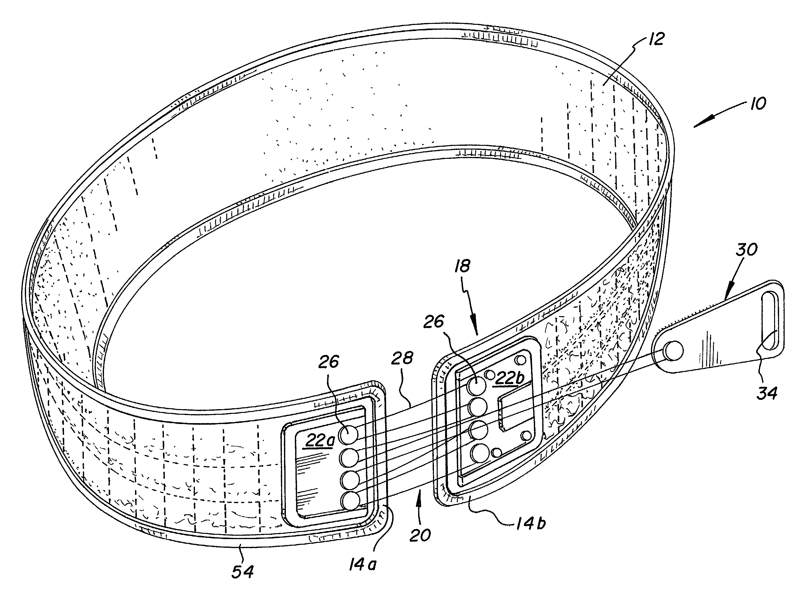
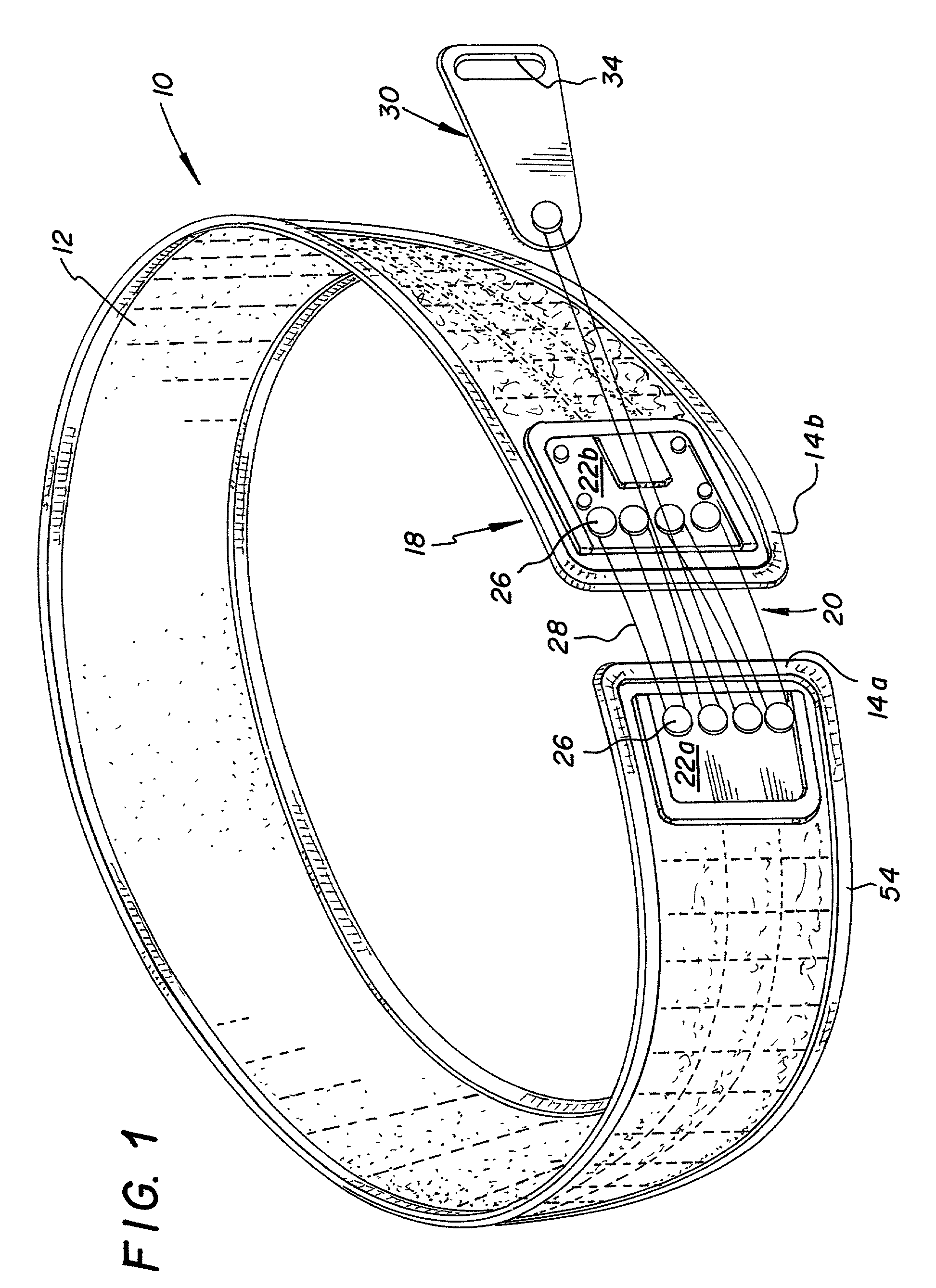
[0020] FIG. 1 shows a first embodiment of the present invention. FIG. 1 illustrates an orthotic trauma device 10 in an assembled position as it would be worn by a patient. The orthosis 10 includes a bracing portion or orthosis body 12, generally having a belt-like shape and made of a material suitable to achieve the type of support sought in treating the condition of the patient wearing the orthotic device. Typically this would include pliant and semi-rigid materials, although a more pliant material is generally preferred to conform substantially to the body of the wearer and to provide support but avoid causing pressure problems to the skin, such as the formation of blisters. The material from which the orthosis body is formed should also be easily cut by a scissors or shears, particularly of the type commonly used or found in hospitals, emergency medical facilities, ambulances, or first aid kits to cut various kinds of adhesive tape and gauze. Suitable materials include canvas, co...


PUM


Abstract

An orthotic trauma device is provided including an elongated orthosis body adapted to be wrapped around a portion of a body of a wearer of the device, the orthosis body being formed of a pliant material which is adapted to be easily cut with cloth-cutting scissors; a detachable fastening device provided at distal ends of the elongated orthosis body to releasably secure the distal ends to one another; and means for adjusting the tightness of the orthosis body operatively associated with the detachable fastening device. A pulley system for use in an orthotic device is also provided including a pair of pulley banks arranged in juxtaposed relationship, a first bank of which is adapted to be detachably disposed on a first distal end of an elongated orthosis body and a second bank of pulleys adapted to be detachably disposed on a second distal end of the elongated orthosis body; and a cable interconnecting the two pulley banks and running through a pulley on each of the pulley banks in alteration, such that shortening of the cable pulling the pulley banks together and tightening the orthotic device with the aid of a mechanical advantage dependent upon the number of pulleys mounted on each of the pulley banks.

Description

[0001] The invention relates to orthotic devices which may be used in the emergency treatment of bone fractures. More particularly, the invention relates to orthotic devices for partial or substantial immobilization of bone fractures, and particularly pelvic fractures, in emergency rooms of hospitals, at an accident site or in transit from an accident site, where quick and effective stabilization of a fracture is essential and where such a device must be adapted both to the dimensions of the individual and the body part being treated.[0002] A common method of treating fractures in the field or at an accident scene is to determine the nature and general extent of the injury and thereafter attempt to immobilize the fractured body part to avoid further injury to the patient, should the body part be moved. Although it may be appropriate to immobilize bone fractures of a limb with a cast arrangement at an accident scene, because of the nature of the type of materials used to form such ca...

Claims


Application Information

Patent Timeline
Patent Type & Authority Applications(United States)
IPC IPC(8): A61F5/03
CPCA61F5/03
Inventor HEINZ, THOMAS J.PARK, DAE SHIKSTEFANSSON, RAFN
Owner PYNG MEDICAL CORP
Why Eureka
Try Eureka