

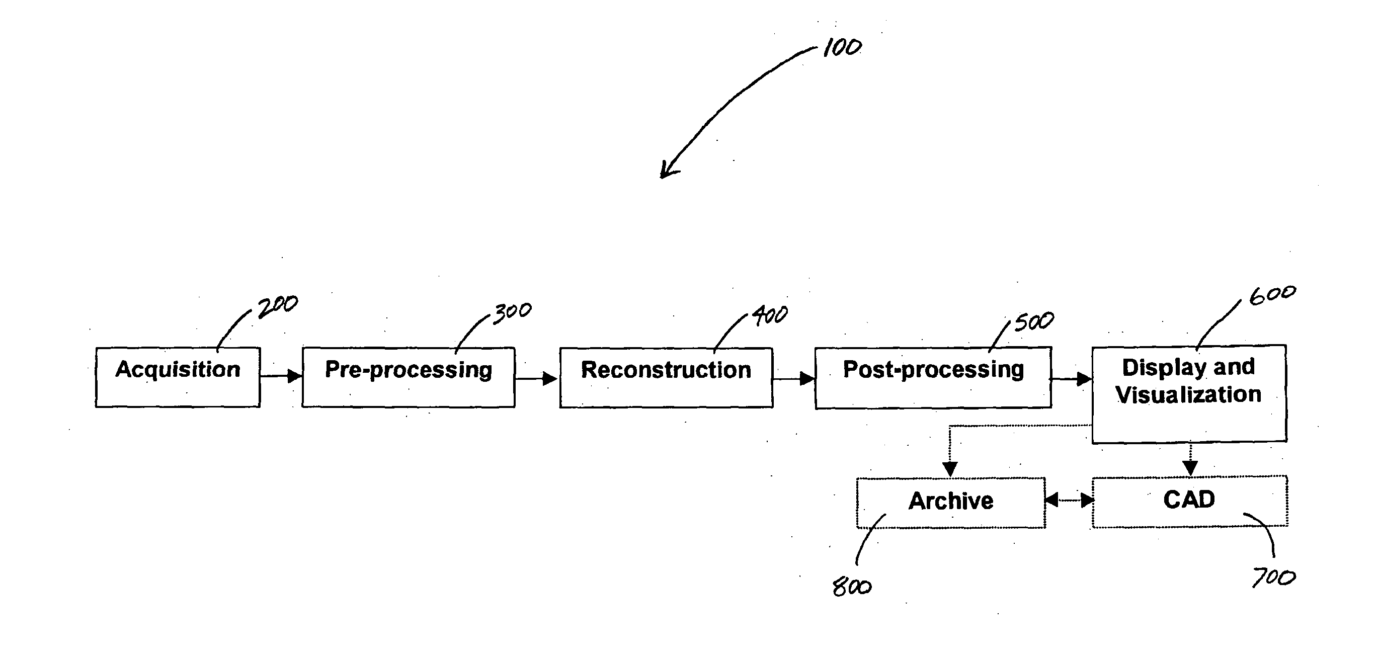
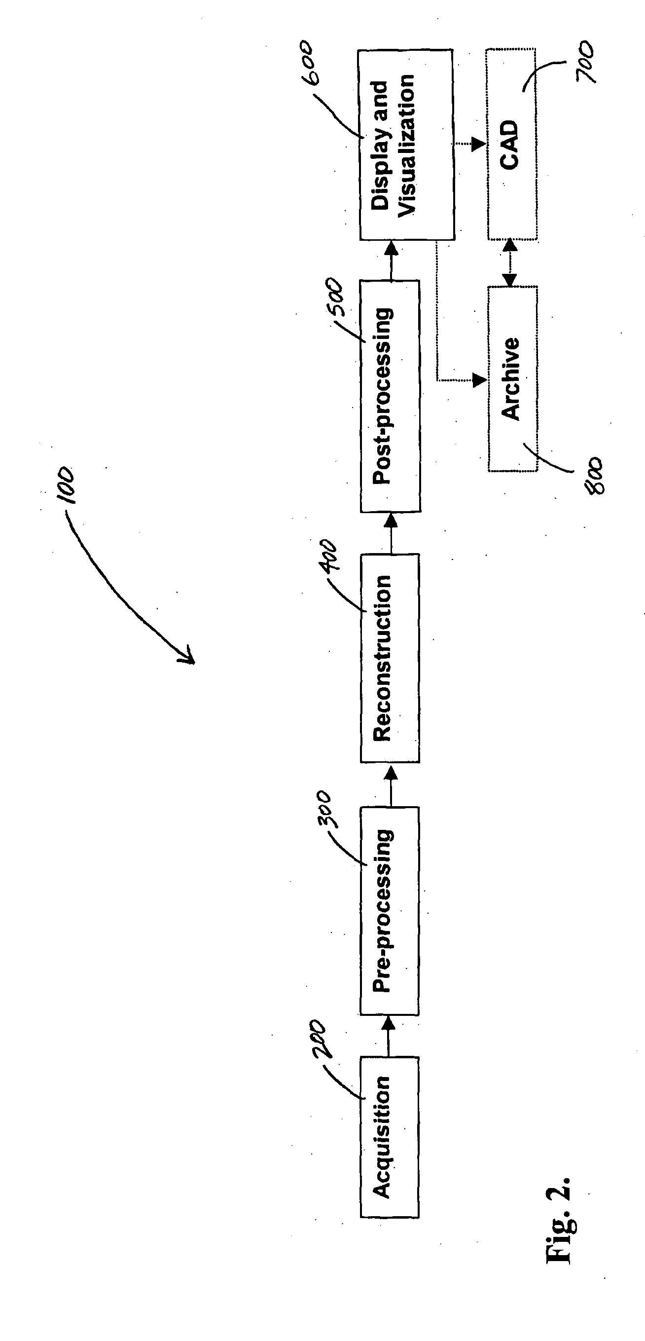
Imaging chain for digital tomosynthesis on a flat panel detector
a detector and digital tomography technology, applied in the field of digital tomography, can solve the problems of image enhancement, image that includes motion artifacts, and images that include motion artifacts, and achieve the effect of enhancing information provided
- Summary
- Abstract
- Description
- Claims
- Application Information
AI Technical Summary
Benefits of technology
Problems solved by technology
Method used
Image

Examples
Embodiment Construction
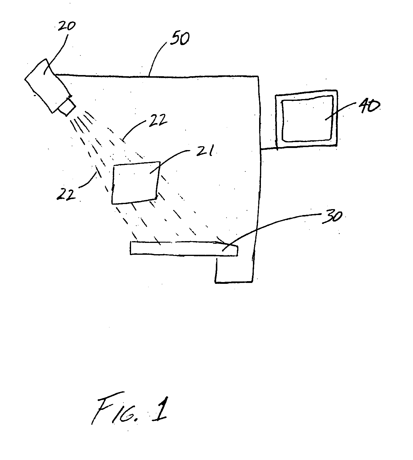
[0018] Referring to FIG. 1, a tomosynthesis system 10 is shown schematically according to a preferred embodiment. Tomosynthesis system 10 includes an x-ray source 20, a detector 30, a computer 40, and supporting structure 50.
[0019] X-ray source 20 is directed toward a subject 21 (e.g., object, patient, etc.) and is configured to emit a beam of x-rays 22 at desired times. Once x-rays 22 are emitted, they pass through subject 21 and are picked up by, or hit, detector 30.
[0020] Detector 30 (e.g., x-ray detector, digital radiography detector, flat panel detector, flat detector, etc.) may be any one of a variety of different detectors conventionally known within the art or that will become available in the future (e.g., energy discriminating detectors that are theoretically capable of acquiring high and low energy images simultaneously). However, according to a preferred embodiment, detector 30 is a flat panel digital detector. When x-rays 22 are picked up by detector 30, they are conv...
PUM
 Abstract
Description
Claims
Application Information