Method for automatically merging a 2D fluoroscopic C-arm image with a preoperative 3D image with one-time use of navigation markers

a technology of navigation markers and c-arms, which is applied in the field of automatic merging of 2d images obtained with carm xray systems, can solve the problem that the anatomy of the patient can only be insufficiently visualized in the 2d fluoroscopic images

Inactive Publication Date: 2005-02-03
SIEMENS AG
View PDF6 Cites 113 Cited by
  • Summary
  • Abstract
  • Description
  • Claims
  • Application Information

AI Technical Summary

Benefits of technology

[0006] An object of the present invention is to merge an intraoperatively obtained 2D fluoroscop

Problems solved by technology

A problem from a medical-technical point of view is that the medical instrument (in the following, a catheter will be referred to as a non-restrictive example) during the procedure (operation or examination) can be visualized very exactly and with high resolutio

Method used


Image

  • Method for automatically merging a 2D fluoroscopic C-arm image with a preoperative 3D image with one-time use of navigation markers
  • Method for automatically merging a 2D fluoroscopic C-arm image with a preoperative 3D image with one-time use of navigation markers
  • Method for automatically merging a 2D fluoroscopic C-arm image with a preoperative 3D image with one-time use of navigation markers

Examples


Embodiment Construction

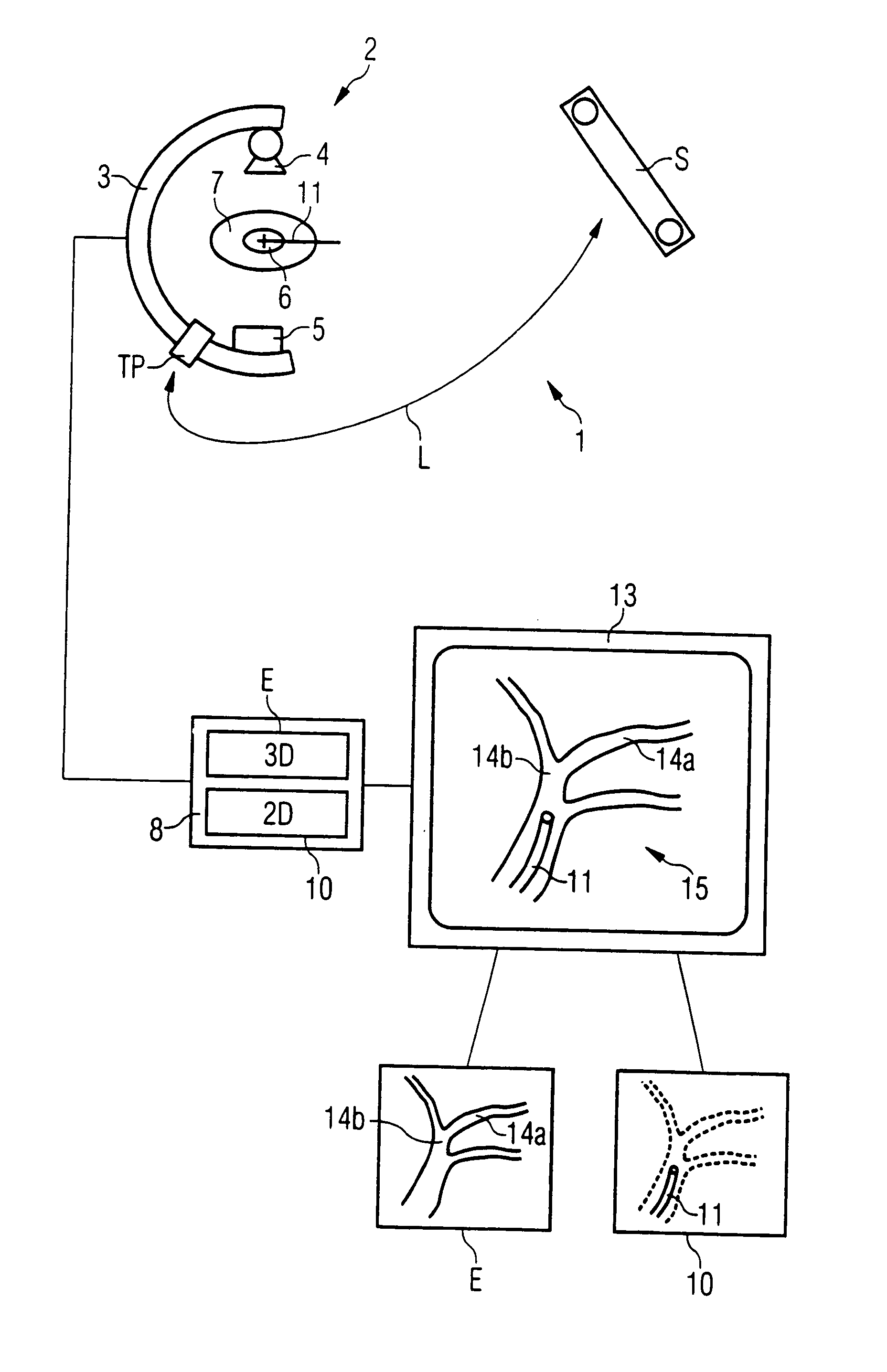
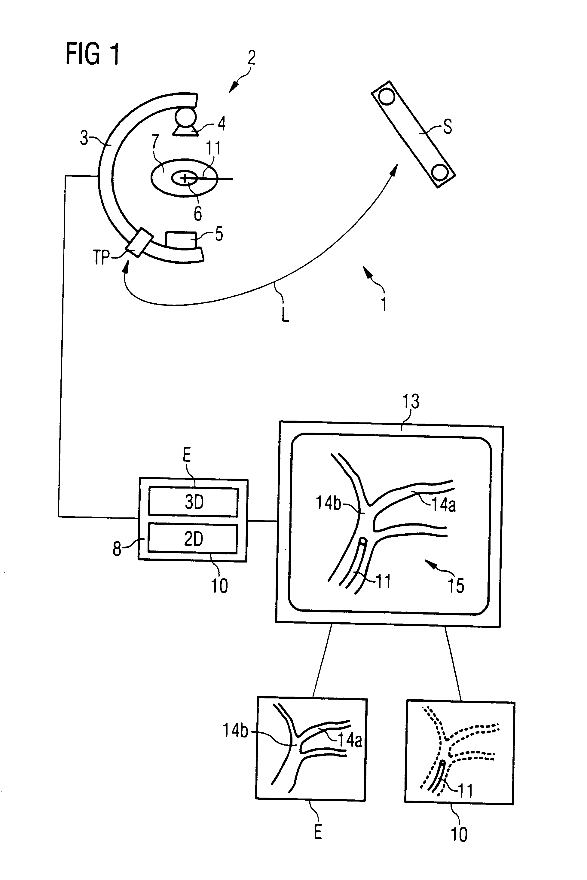
[0018]FIG. 1 schematically illustrates an examination and / or treatment system 1 in accordance with the invention, with only basic components being shown. The system includes an imaging system 2 to obtain two-dimensional transillumination images (2D fluoroscopic images). The imaging system 2 has a C-arm 3, to which an X-ray radiation source 4, and a radiation detector 5, for instance a solid body imaging detector, and a tool plate TP are attached. The examination zone 6 of a patient 7 is located ideally in the isocenter of the C-arm 3, so that its entire extent is visible in the captured 2D fluoroscopic image.

[0019] In the immediate vicinity of the imaging system 2, there is a navigation sensor S, by means of which the current position of the tool plate TP can be recorded and thus the C-arm 3, as well as the position and orientation of a medical instrument 11 used for the procedure, and the patient.

[0020] The system 1 is operated using a control and processing unit 8, which among o...


PUM


Abstract

In a method and apparatus for the automatic merging of 2D fluoroscopic C-arm images with preoperative 3D images with a one-time use of navigation markers, markers in a marker-containing preoperative 3D image are registered relative to a navigation system, a tool plate fixed on the C-arm system is registered in a reference position relative to the navigation system, a 2D C-arm image (2D fluoroscopic image) that contains the image of at least a medical instrument is obtained in an arbitrary C-arm position, a projection matrix for a 2D-3D merge is determined on the basis of the tool plate and the reference position relative to the navigation system, and the 2D fluoroscopic image is superimposed with the 3D image on the basis of the projection matrix.

Description

BACKGROUND OF THE INVENTION [0001] 1. Field of the Invention [0002] The present invention concerns a method for superimposing or fusing a 2D image obtained with a C-arm x-ray system, with a preoperative 3D image. The invention particularly concerns the display of a medical instrument in the 3D image which is in the examination region of a patient and is included in the 2D image. [0003] 2. Description of the Prior Art [0004] An increasing number of examinations or treatments of patients are performed minimally invasively, that is, with the least possible surgical trauma. Examples are treatments with endoscopes, laparoscopes, or catheters, all of which are inserted into the examination zone of a patient through a small opening in the body. Catheters, for example, are often used in the course of cardiological examinations. [0005] A problem from a medical-technical point of view is that the medical instrument (in the following, a catheter will be referred to as a non-restrictive example...

Claims


Application Information

Patent Timeline
IPC IPC(8): A61B5/05A61B19/00G06T7/00G06T17/00
CPCA61B2019/5238A61B2019/5289A61B2019/5291G06T7/0028A61B19/5244G06T2207/30004A61B6/12A61B6/4441G06T7/0038A61B34/20A61B2090/364A61B2090/376A61B2090/365G06T7/33G06T7/38
Inventor MITSCHKE, MATTHIASRAHN, NORBERTRITTER, DIETER
Owner SIEMENS AG
Why Eureka
Try Eureka