
Methods and devices useful for analyzing color medical images

a color medical image and color technology, applied in the field of medical imaging, can solve the problems of difficult to accurately describe colors in device-dependent rgb, the use of device-dependent red-green-blue (rgb) color spaces is a problem, and the transmission of medical color images such as endoscopic and dermatological images has not been discussed intensively

Inactive Publication Date: 2005-02-17
UNIV OF IOWA RES FOUND
View PDF19 Cites 107 Cited by
  • Summary
  • Abstract
  • Description
  • Claims
  • Application Information

AI Technical Summary

Benefits of technology

The invention is a method for comparing a color medical image to a normal color medical image database to identify abnormal pixels. The method involves converting the color medical image to a different color format, such as RGB or HSI, and comparing it to the normal image data. The method can be implemented using a computer system or a device with a processor, such as an FPGA or ASIC. The technical effect of the invention is to provide a reliable and efficient method for identifying abnormal pixels in color medical images, which can aid in the diagnosis and treatment of medical conditions.

Problems solved by technology

The problems concerning the transmission of medical color images such as endoscopic and dermatological images have not, however, been discussed intensively.
Considering that each imaging system has its own time-varying RGB color space that depends on its own unique spectral sensitivities, it remains difficult to accurately describe colors in device-dependent RGB.
Therefore, it has been found that the use of a device-dependent red-green-blue (RGB) color spaces is a problem.

Method used


Image

  • Methods and devices useful for analyzing color medical images
  • Methods and devices useful for analyzing color medical images
  • Methods and devices useful for analyzing color medical images

Examples


Embodiment Construction

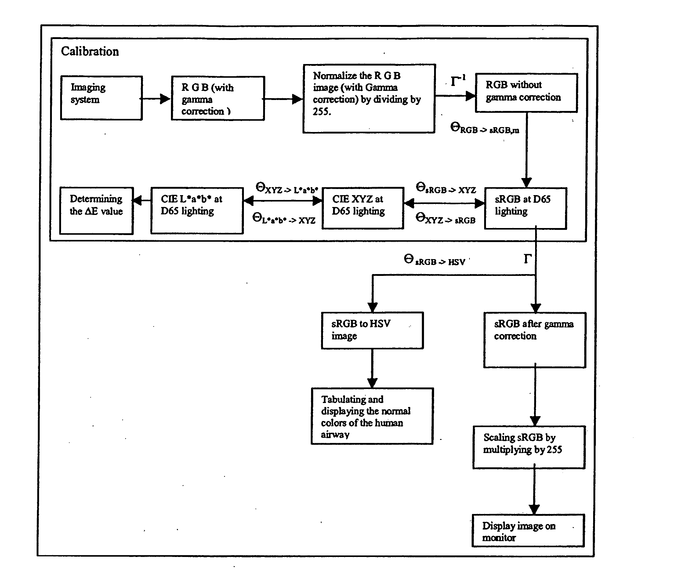
The terms “comprise” (and any form of comprise, such as “comprises” and “comprising”), “have” (and any form of have, such as “has” and “having”), and “include” (and any form of include, such as “includes” and “including”) are open-ended linking verbs. As a result, a method or a device (e.g., a computer readable medium or a computer chip) that “comprises,”“has,” or “includes” one or more steps or elements possesses those one or more steps or elements, but is not limited to possessing only those one or more elements or steps. Thus, a method “comprising” comparing a subject color medical image to normal color medical image data; and identifying abnormal pixels from the subject color medical image is a method that includes these two steps, but is not limited to possessing only the two recited steps. For example, the method also covers methods with the recited two steps and additional steps such as displaying a histogram that includes (i) saturation information about the subject color m...


PUM


Abstract

In one embodiment, a method that includes comparing a subject color medical image to normal color medical image data; and identifying abnormal pixels from the subject color medical image. Another embodiment includes a computer readable medium comprising machine readable instructions for implementing one or more steps of that method. Another embodiment includes a device that has a field programmable gate array configured to perform one or more of the steps of that method. Another embodiment includes a device that has an application specific integrated circuit configured to perform one or more of the steps of that method.

Description

BACKGROUND OF THE INVENTION 1. Field of the Invention The invention relates generally to the field of medical imaging. More particularly, the invention relates to the analysis of color medical images to detect abnormalities in the subject tissue. 2. Discussion of the Related Art Despite the dramatic progress of multimedia information technology and its rapid spread into the medical profession, discussions on medical images so far have concentrated largely on sufficient spatial sampling rate and sufficient grayscale gradations for black and white pictures such as X-ray, CT and MRI. The problems concerning the transmission of medical color images such as endoscopic and dermatological images have not, however, been discussed intensively. Color image analysis has been extensively used in dermatology and in the assessment of wound healing (Herbin et al., Haeghen et al.). Nischik et al. in 1997 developed a method to analyze the spreading of skin erythemas by determining the change in...

Claims


Application Information

Patent Timeline
Patent Type & Authority Applications(United States)
IPC IPC(8): G06T7/00G06T7/40G06V10/56
CPCG06K9/4652G06T7/0012G06T2207/30004G06T2200/24G06T2207/10016G06T7/408G06T7/90G06V10/56
Inventor MCLENNAN, GEOFFREYDONNELLEY, MARTINGOPALAKRISHNAN, DEEPAHOFFMAN, ERICREINHARDT, JOSEPHSUTER, MELISSA
Owner UNIV OF IOWA RES FOUND
Learn More