
Intracorneal lens placement method and apparatus

a lens placement and intracorneal technology, applied in the field of ophthalmologic surgery, can solve the problems of difficult surgery to accurately perform, unsatisfactory lenses for such vision correction, and risk of creating permanent visual aberrations for patients, so as to facilitate the retention of the lens, improve the amplitude of the lateral oscillation, and minimize the disturbance of the surgical setup

Inactive Publication Date: 2005-03-03
NEOPTICS AG
View PDF10 Cites 31 Cited by
  • Summary
  • Abstract
  • Description
  • Claims
  • Application Information

AI Technical Summary

Benefits of technology

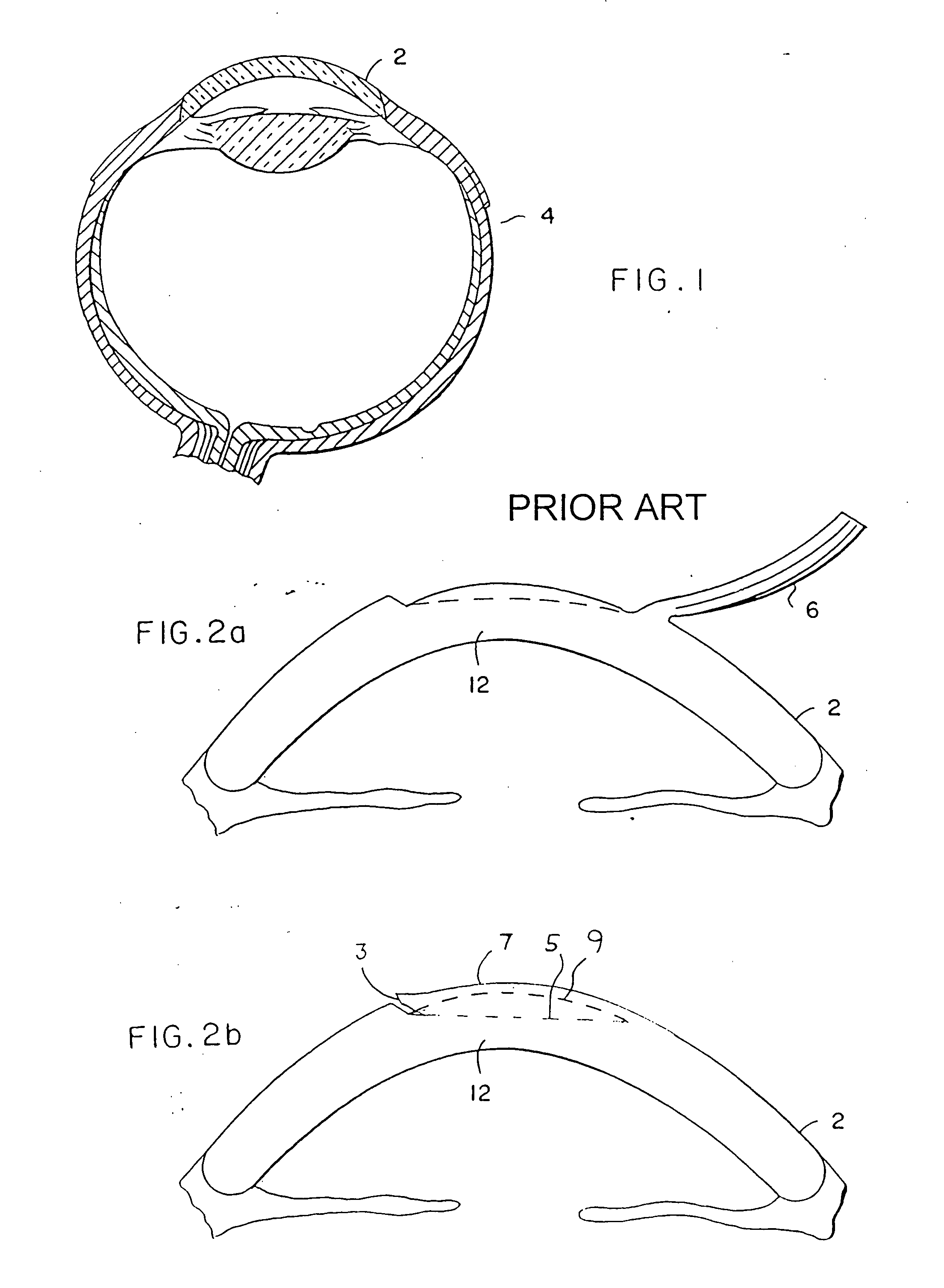
[0007] The present invention solves the above-noted need by providing a method and devices for intracorneal lens placement. A specially adapted lens is implanted in a corneal pocket which has been precisely formed by a device which creates and shapes the pocket to accept and retain a lens in the cornea. Whereas in typical corrective surgery an entire flap of the cornea is lifted as shown in FIG. 2a to permit access for further surgical modification of the cornea, in vision modification according to the present invention a flap of cornea is not lifted, but rather a pocket is formed in the corneal tissue as shown in FIG. 2b. As much of the corneal surface as practical is left intact to simplify healing and to discourage movement or loss of the inserted lens.
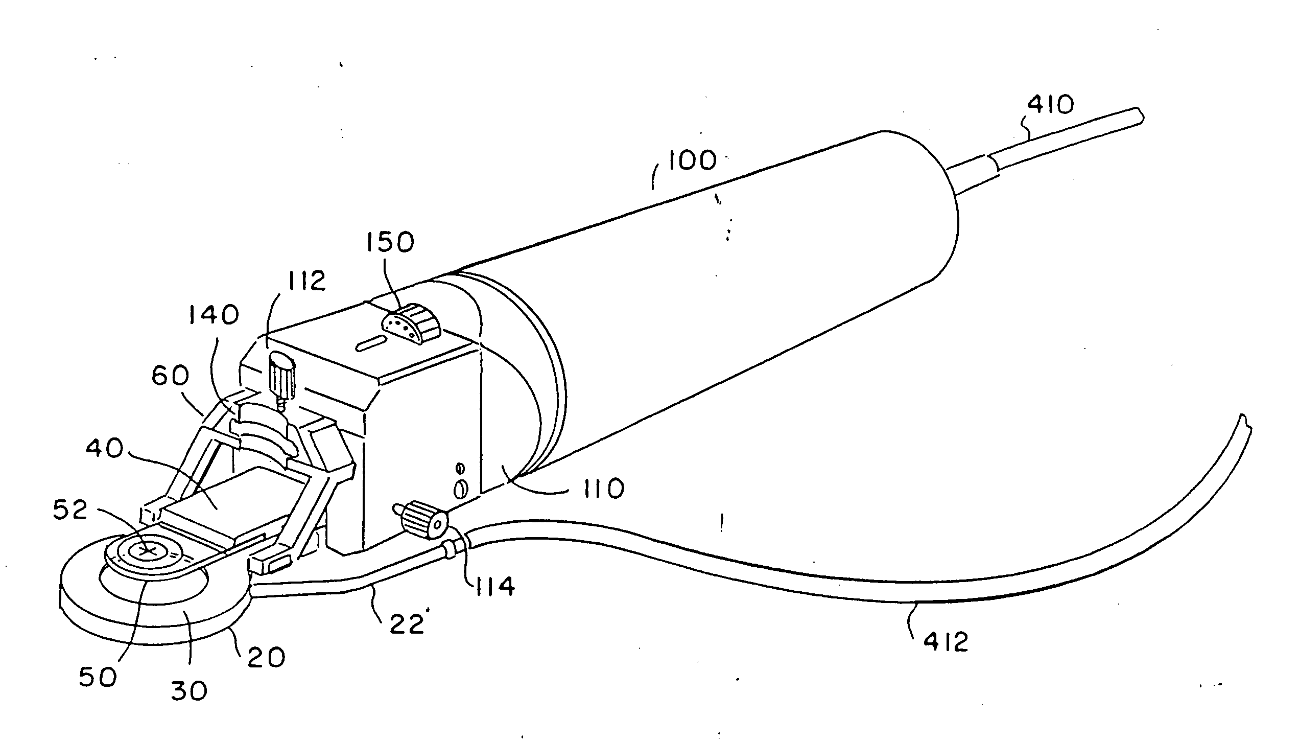
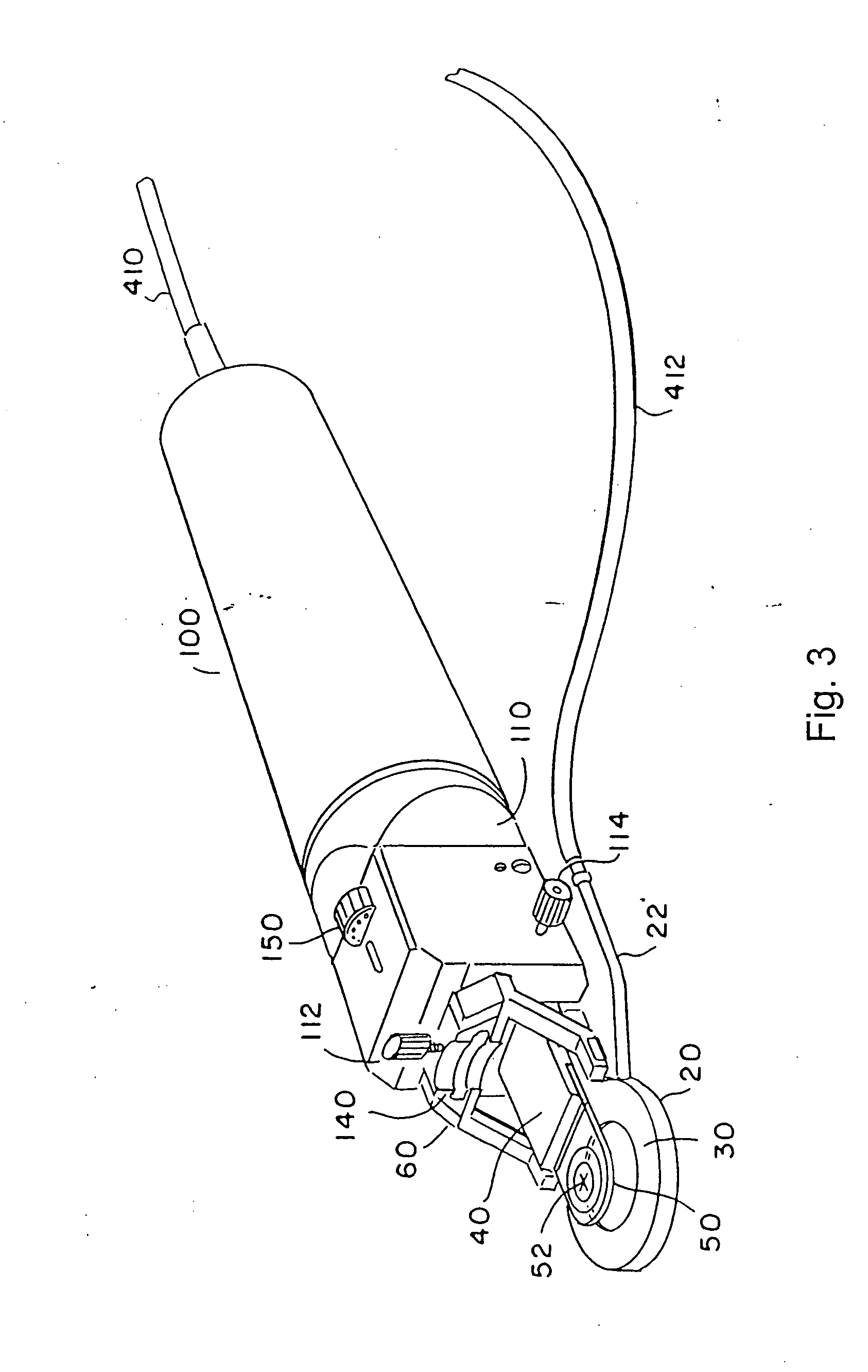
[0008] In order to position a lens within the cornea of an eye in a precisely predictable and repeatable manner, and to help retain the intended orientation and positioning of the lens while the eye heals from surgery, the present invention provides a corneal pocket keratome to create a pocket of precise dimensions in the cornea, and also a lens having special features to establish a close fit between the lens and the corneal pocket. Both of these pieces can be realized in a number of different embodiments. Moreover, the corneal pocket keratome has several subparts, each of which can be realized in many ways.
[0009] The lens size and shape matches the corneal pocket formed by the corneal pocket keratome, and provides desired focal modifications when disposed within corneal tissue. The lens permits sufficient gas diffusion to allow adequate oxygenation of internal eye tissues. In preferred embodiments, lens features create an interference fit between the lens and the corneal tissue at the edges of the corneal pocket to aid in retaining the placement and orientation of the lens. In addition to a precise fit, such retention features of the lens may include a material which swells when hydrated after placement within the cornea, or variations in the radius of the lens to form circumferential bumps. The lens may accordingly have an asymmetric, radially and / or axially varying focus to compensate for the effects of astigmatism or presbyopia, generally in addition to compensation for myopia or hyperopia. For some applications, lens thickness may be desirably reduced by employing a Fresnel intracorneal lens.

Problems solved by technology

However, most such surgical reshaping is not reversible, resulting in some risk of creating permanent visual aberrations for the patient.
Such surgery is difficult to perform accurately.
Moreover, the lenses which are available for such vision correction are not entirely satisfactory for a variety of reasons, including a tendency to shift out of position after placement, to impair transcorneal gas diffusion, to be excessively thick, or to be unable to correct presbyopia or astigmatism.

Method used


Image

  • Intracorneal lens placement method and apparatus
  • Intracorneal lens placement method and apparatus
  • Intracorneal lens placement method and apparatus

Examples


first embodiment

A First Embodiment for a Pocket Keratome

[0132] In a further development the invention is a pocket keratome that will cut a pocket in the cornea into which a lens or other device can be inserted.

[0133] The pocket keratome has a blade support and guide member constructed as a unitary part to mount on a drive mechanism of the type described above that can drive it forward while reciprocating laterally. The term unitary part as used herein means that it is made from one piece of material, preferably stainless steel. In particular the blade support and guide member are made from one piece. The blade is mounted on the blade support for precise positioning with respect to the guide. Also a positioning ring as described above is provided.

[0134] In accordance with a major goal of a pocket keratome, the blade support and guide member is configured such that when assembled with the blade, it will enable a very precise and precisely controllable depth of cut in the cornea. The depth of cut is...


PUM


Abstract

A corneal-pocket keratome having a support and blade assembly and in which the support has a guide on a forward portion in front of the blade and spaced precisely from the blade to control entry of the blade into the cornea to precisely position the pocket in the cornea that is cut by the blade. A lens is placed in the pocket.

Description

[0001] The present application is a continuation in part of copending U.S. patent application Ser. No. 09 / 132,987, filed Aug. 12, 1998, which is incorporated herein by reference.FIELD OF THE INVENTION [0002] The present invention pertains to the general field of ophthalmologic surgery, and in particular to surgical methods and devices for corneal implantation of optical lenses. BACKGROUND [0003] Numerous ophthalmic surgical procedures have been developed for correcting imperfect visual acuity such as myopia or hyperopia. A variety of keratomes have been developed over recent decades, devices for performing corneal resectioning to permit access to inner portions of the cornea, where surgical reshaping may then be used to permanently correct vision defects. [0004] Referring to FIGS. 1 and 2a, a typical prior art resectioning operation will separate flap 6 of corneal (and epithelial) tissue 2 from eyeball 4. The outer layers of cornea and epithelial cells are separated and lifted away ...

Claims


Application Information

Patent Timeline
Patent Type & Authority Applications(United States)
IPC IPC(8): A61F9/013
CPCA61F9/013
Inventor FEINGOLD, VLADIMIRLOSMYNINE, ALEX
Owner NEOPTICS AG
Learn More