Eureka AIR delivers breakthrough ideas for toughest innovation challenges, trusted by R&D personnel around the world.

Intracorneal lens placement method and apparatus

a technology of intracorneal lens and placement method, which is applied in the field of ophthalmologic surgery, can solve the problems of difficult surgery, insufficient lens availability for such vision correction, and risk of creating permanent visual aberrations for patients, so as to facilitate the retention of the lens, improve the amplitude of the lateral oscillation, and minimize the disturbance of the surgical setup

Inactive Publication Date: 2007-04-24
BIOVISION AG
View PDF2 Cites 49 Cited by
  • Summary
  • Abstract
  • Description
  • Claims
  • Application Information

AI Technical Summary

Benefits of technology

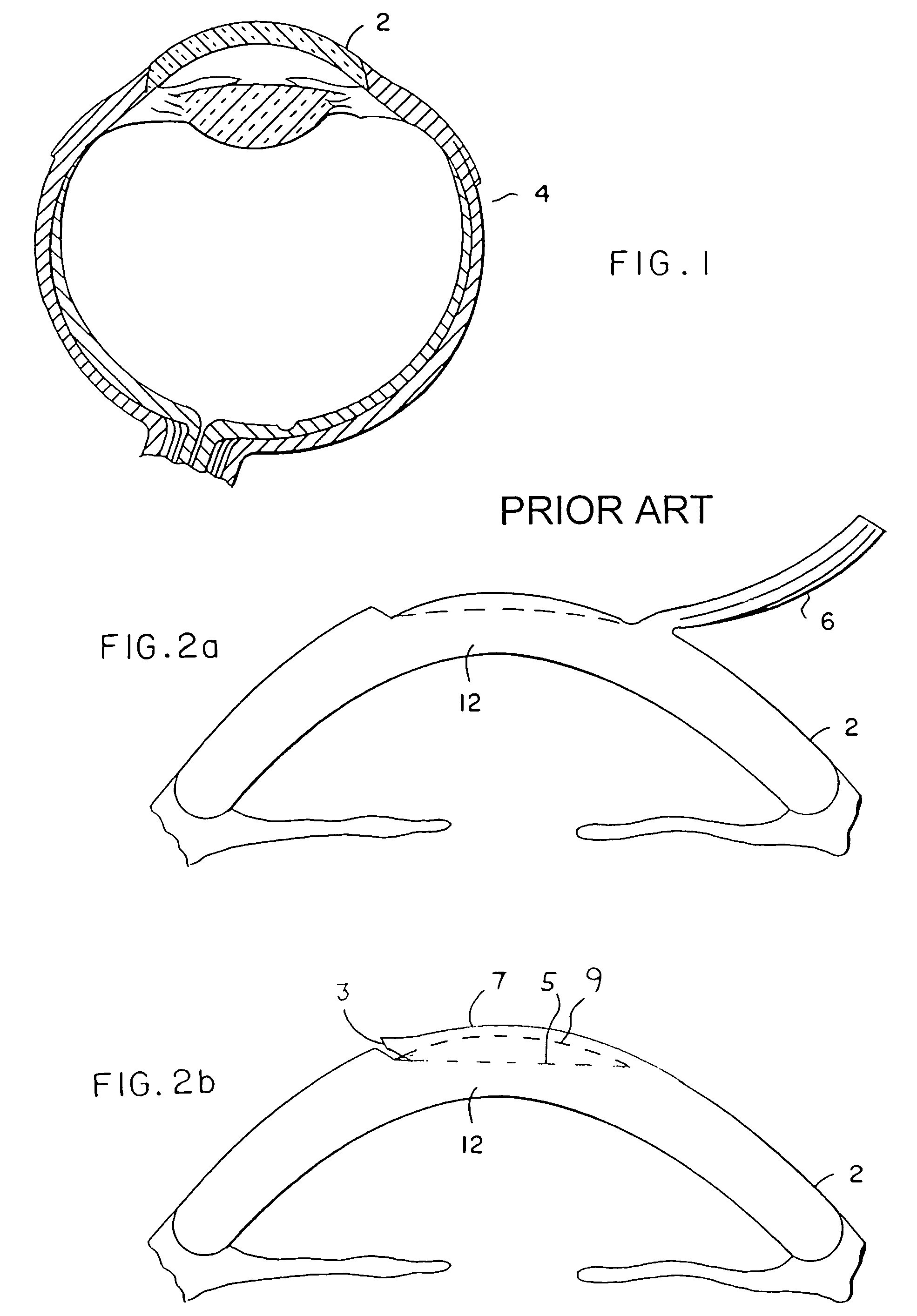
[0007]The present invention solves the above-noted need by providing a method and devices for intracorneal lens placement. A specially adapted lens is implanted in a corneal pocket which has been precisely formed by a device which creates and shapes the pocket to accept and retain a lens in the cornea. Whereas in typical corrective surgery an entire flap of the cornea is lifted as shown in FIG. 2a to permit access for further surgical modification of the cornea, in vision modification according to the present invention a flap of cornea is not lifted, but rather a pocket is formed in the corneal tissue as shown in FIG. 2b. As much of the corneal surface as practical is left intact to simplify healing and to discourage movement or loss of the inserted lens.
[0008]In order to position a lens within the cornea of an eye in a precisely predictable and repeatable manner, and to help retain the intended orientation and positioning of the lens while the eye heals from surgery, the present invention provides a corneal pocket keratome to create a pocket of precise dimensions in the cornea, and also a lens having special features to establish a close fit between the lens and the corneal pocket. Both of these pieces can be realized in a number of different embodiments. Moreover, the corneal pocket keratome has several subparts, each of which can be realized in many ways.
[0009]The lens size and shape matches the corneal pocket formed by the corneal pocket keratome, and provides desired focal modifications when disposed within corneal tissue. The lens permits sufficient gas diffusion to allow adequate oxygenation of internal eye tissues. In preferred embodiments, lens features create an interference fit between the lens and the corneal tissue at the edges of the corneal pocket to aid in retaining the placement and orientation of the lens. In addition to a precise fit, such retention features of the lens may include a material which swells when hydrated after placement within the cornea, or variations in the radius of the lens to form circumferential bumps. The lens may accordingly have an asymmetric, radially and / or axially varying focus to compensate for the effects of astigmatism or presbyopia, generally in addition to compensation for myopia or hyperopia. For some applications, lens thickness may be desirably reduced by employing a Fresnel intracorneal lens.

Problems solved by technology

However, most such surgical reshaping is not reversible, resulting in some risk of creating permanent visual aberrations for the patient.
Such surgery is difficult to perform accurately.
Moreover, the lenses which are available for such vision correction are not entirely satisfactory for a variety of reasons, including a tendency to shift out of position after placement, to impair transcorneal gas diffusion, to be excessively thick, or to be unable to correct presbyopia or astigmatism.

Method used


Image

  • Intracorneal lens placement method and apparatus
  • Intracorneal lens placement method and apparatus
  • Intracorneal lens placement method and apparatus

Examples


Embodiment Construction

[0044]The present invention presents means to permanently, yet reversibly, correct defects of vision by disposing a lens in a pocket in a cornea. Various embodiments correct myopia, hyperopia, astigmatism, presbyopia, or a combination of these defects. Appropriate lenses are provided, as well as a device to create a corneal pocket to accept these lenses. The correction may be permanent, if it remains satisfactory, and may also be reversed by removing the lens from the cornea.

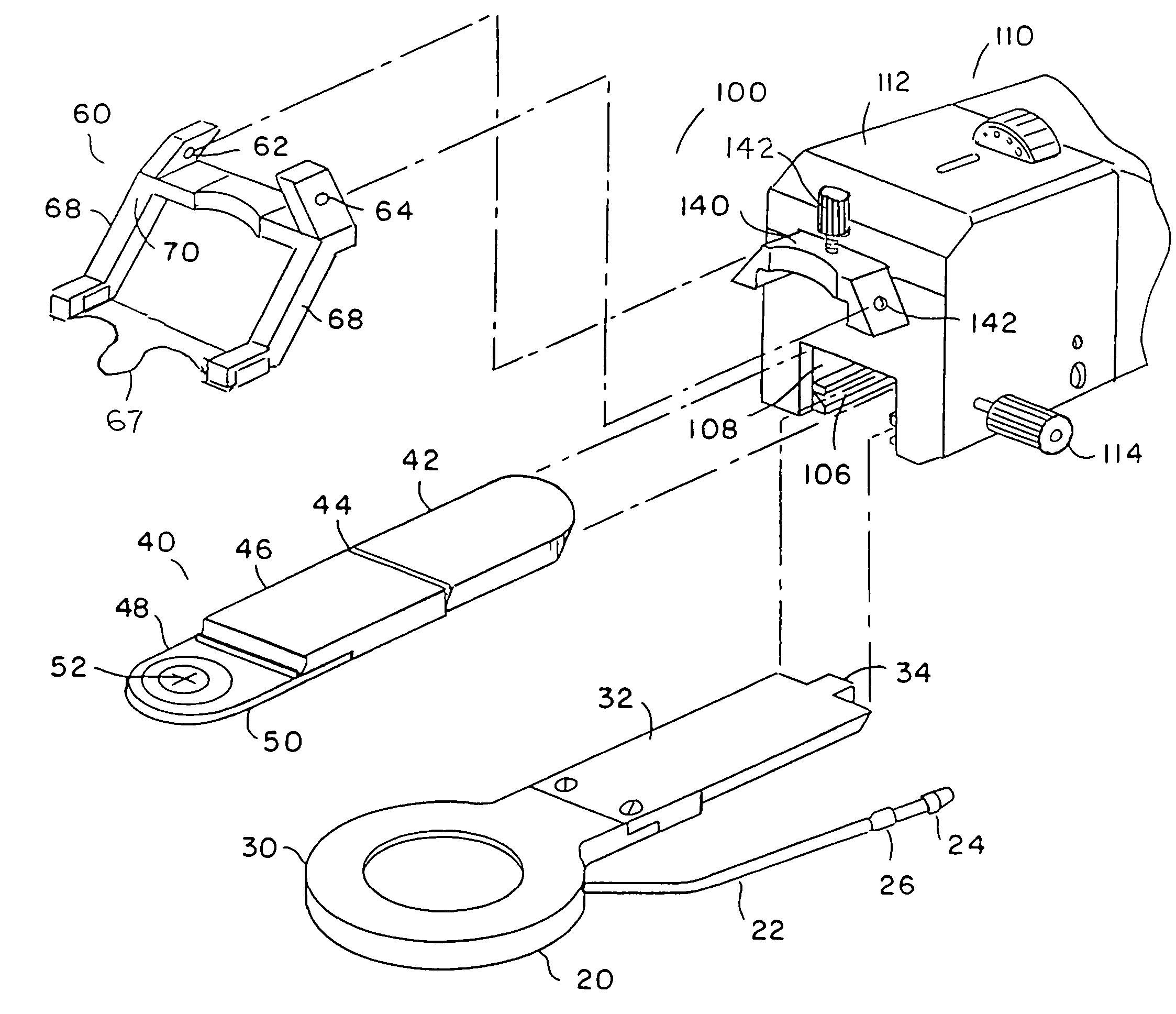
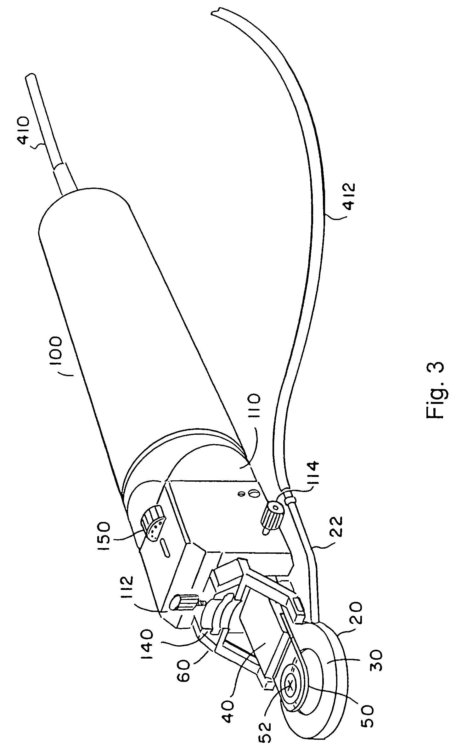
[0045]We begin with an overview of a device for preparing a corneal pocket to retain an appropriate lens in a subject eye. Referring to FIGS. 3, 4 and 5, such a device is preferably embodied in three separate components: surgical unit 100, footpedal 300, and control unit 400. Surgical unit 100 has four subsections including drive assembly 110 and three cutting head elements: positioning ring assembly 20, optional applanator assembly 40, and blade fork assembly 60. Footpedal 300 communicates user commands to cont...


PUM


Abstract

A method and apparatus for correcting vision, including a corneal-pocket keratome device to create a corneal pocket and a lens to be inserted and retained in the corneal pocket to effect correction. The corneal-pocket keratome includes a drive unit having cutting head elements which contact the subject eye during corneal pocket formation. The cutting head elements may be removeable and may be disposable. The cutting head elements include a corneal restraint device, which may be a positioning ring to position an eyeball with the cornea protruding through the ring; a keratome blade assembly with a corneal-pocket blade; and may also include an applanation shoe surface to restrain the cornea, in addition or instead of the positioning ring. The applanation shoe may be pivotable away from the surgical area. The corneal-pocket blade may include a guide which travels with the blade. The blade assembly oscillates laterally while extending forward into the cornea to form the pocket, and the amplitude of the lateral oscillation is preferably increased as the blade goes beyond an opening incision into the cornea. Lenses for this invention preferably include a feature to impede accidental lens movement after the lens is disposed within the corneal pocket, which may be a swelling after insertion or a circumferential irregularity. Lenses may be of Fresnel or non-Fresnel type, and may employ annular changes in the index of refraction of the lens material, as well as changes in refractive shape which may be annular or not, to effect variations in focal length for relieving presbyopia, astigmatism, and combinations of those as well as myopia and hyperopia. Drive control and vacuum for the positioning ring are provided under user command by a control unit having user inputs.

Description

[0001]The present application is a divisional of application Ser. No. 09 / 586,273 filed on Jun. 2, 2000 now U.S. Pat. No. 6,599,305 which is a continuation-in-part of application Ser. No. 09 / 132,987 filed on Aug. 12, 1998 now U.S. Pat. No. 6,083,236 issued on Jul. 4, 2000 the contents of all of which are incorporated herein by reference.FIELD OF THE INVENTION[0002]The present invention pertains to the general field of ophthalmologic surgery, and in particular to surgical methods and devices for corneal implantation of optical lenses.BACKGROUND[0003]Numerous ophthalmic surgical procedures have been developed for correcting imperfect visual acuity such as myopia or hyperopia. A variety of keratomes have been developed over recent decades, devices for performing corneal resectioning to permit access to inner portions of the cornea, where surgical reshaping may then be used to permanently correct vision defects.[0004]Referring to FIGS. 1 and 2a, a typical prior art resectioning operation...

Claims


Application Information

Patent Timeline
Patent Type & Authority Patents(United States)
IPC IPC(8): A61F9/00A61B17/32A61F2/14A61F2/16A61F9/007A61F9/013
CPCA61F9/013A61F9/0133A61F2/1451
Inventor FEINGOLD, VLADIMIR
Owner BIOVISION AG
Why Eureka
Eureka Blog
Learn More