Method and apparatus for measuring bladder electrical activity to diagnose bladder dysfunction

a technology of electrical activity and bladder, applied in the field of methods and apparatus to diagnose bladder pathology from surface electrode potential, can solve the problems of patient discomfort, inability to perform ambulatory setting, invasiveness of urodynamic tests,

Inactive Publication Date: 2005-03-17
BERGER YITZHAK +1
View PDF2 Cites 21 Cited by
  • Summary
  • Abstract
  • Description
  • Claims
  • Application Information

AI Technical Summary

Problems solved by technology

In addition, the urodynamic tests are invasive in nature, and they cannot be performed in an ambulatory setting.
Urodynamic testing must b...

Method used


Image

  • Method and apparatus for measuring bladder electrical activity to diagnose bladder dysfunction
  • Method and apparatus for measuring bladder electrical activity to diagnose bladder dysfunction
  • Method and apparatus for measuring bladder electrical activity to diagnose bladder dysfunction

Examples


Embodiment Construction

[0028] The description is divided into two parts. Part I introduces the inventive methods and apparatus and explains the significance of the invention as a new diagnostic technique in urology. Part II presents the apparatus in more detail, including engineering aspects related to the hardware and software. Appendix I describes additional exemplary display screens according to one embodiment of the apparatus.

PART I: BLADDER SPHINCTER AND DETRUSOR MUSCLE VISEMG

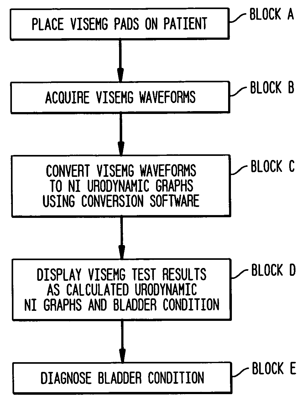
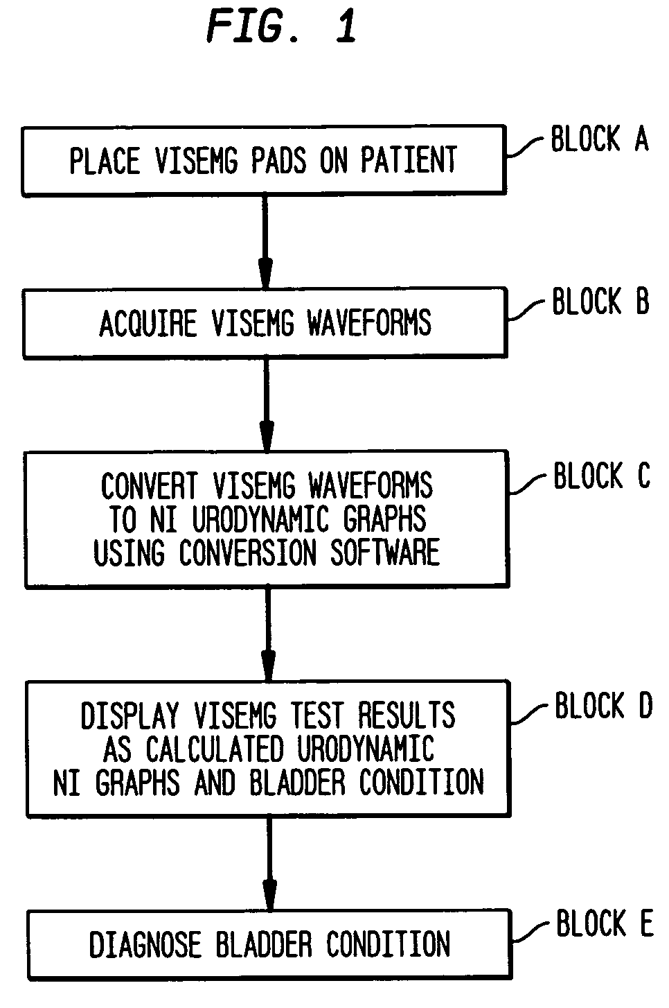
[0029] A first embodiment of the inventive method is shown in FIG. 1. In this non-invasive diagnostic test, Vesico Internal Sphincter ElectroMyogram (VISEMG) electrodes are placed on the patient (Block A). The VISEMG electrodes are surface electrodes that record electromyographic signals from the bladder and internal sphincter muscles. The VISEMG waveforms are then acquired from the electrodes (Block B).

[0030] The VISEMG waveforms are converted to non-invasive (NI) urodynamic graphs (Block C). NI urodynamic graphs are graphs ...


PUM


Abstract

A method of diagnosing bladder pathology uses Vesico Internal Sphincter ElectroMyogram (VISEMG) waveforms obtained from a plurality of VISEMG electrodes disposed on a patient. The VISEMG waveforms are converted to noninvasive (NI) urodynamic graphs. Further, a programmed computer assesses the condition of the bladder based on the VISEMG waveforms. The method does not require traditional urodynamics or any other invasive procedure. An apparatus to perform the inventive method uses a plurality of VISEMG electrodes placed on the patient. Each electrode signal is amplified and filtered and converted to a digital signal. A computer converts the digital signals (the VISEMG waveforms) to NI urodynamic graphs. Any combination of the VISEMG waveforms, the NI urodynamic graphs, and the condition of the bladder based on the VISEMG waveforms can then be shown on a display screen.

Description

FIELD OF THE INVENTION [0001] This invention relates to a method and apparatus to diagnose bladder pathology from surface electrode potentials, and more specifically to a method and apparatus to convert surface potential measurements into graphs similar to those obtained by conventional invasive urodynamic testing. The resulting non-invasive urodynamic graphs can then be used to diagnose bladder pathology. BACKGROUND OF THE INVENTION [0002] The bladder collects and stores urine from the kidneys and allows for discharge of the urine. The two bladder functions are controlled by the sympathetic and the parasympathetic autonomic neurological systems, and these are coordinated by the brain via the spinal cord. [0003] The first function is to store urine in a low pressure environment without leakage. The detrusor, or bladder muscle, maintains low pressure within the bladder cavity during the storage phase. A sympathetic input prevents an increase of intravesical pressure during the accumu...

Claims


Application Information

Patent Timeline
IPC IPC(8): A61B5/0488
CPCA61B5/726A61B5/04882A61B5/391
Inventor BERGER, YITZHAKBALDAR, ISSAC P.
Owner BERGER YITZHAK
Why Eureka
Try Eureka