Method and apparatus for single transmission Golay coded excitation

a single-transmission, coded excitation technology, applied in the field of ultrasound imaging, can solve the problems of preventing reliable analysis of images, poor image quality, and reducing image contrast quality, and achieve the effect of improving ultrasound imaging

Inactive Publication Date: 2005-05-05
GE MEDICAL SYST GLOBAL TECH CO LLC
View PDF20 Cites 29 Cited by
  • Summary
  • Abstract
  • Description
  • Claims
  • Application Information

AI Technical Summary

Problems solved by technology

Poor image quality may prevent reliable analysis of an image.
For example, a decrease in image contrast quality may yield an unreliable image that is not usable clinically.
Poor resolution introduces additional noise or interference in an image.
However, increasing transmitted amplitude of a signal has limitations related to patient safety, maximum drive levels, and nonlinear distortions.
However, increasing pulse duration has the undesirable consequence of reducing axial resolution.
However, Golay coded excitation is rarely used in current systems due to increased transmission and acquisition time.

Method used


Image

  • Method and apparatus for single transmission Golay coded excitation
  • Method and apparatus for single transmission Golay coded excitation
  • Method and apparatus for single transmission Golay coded excitation

Examples


Embodiment Construction

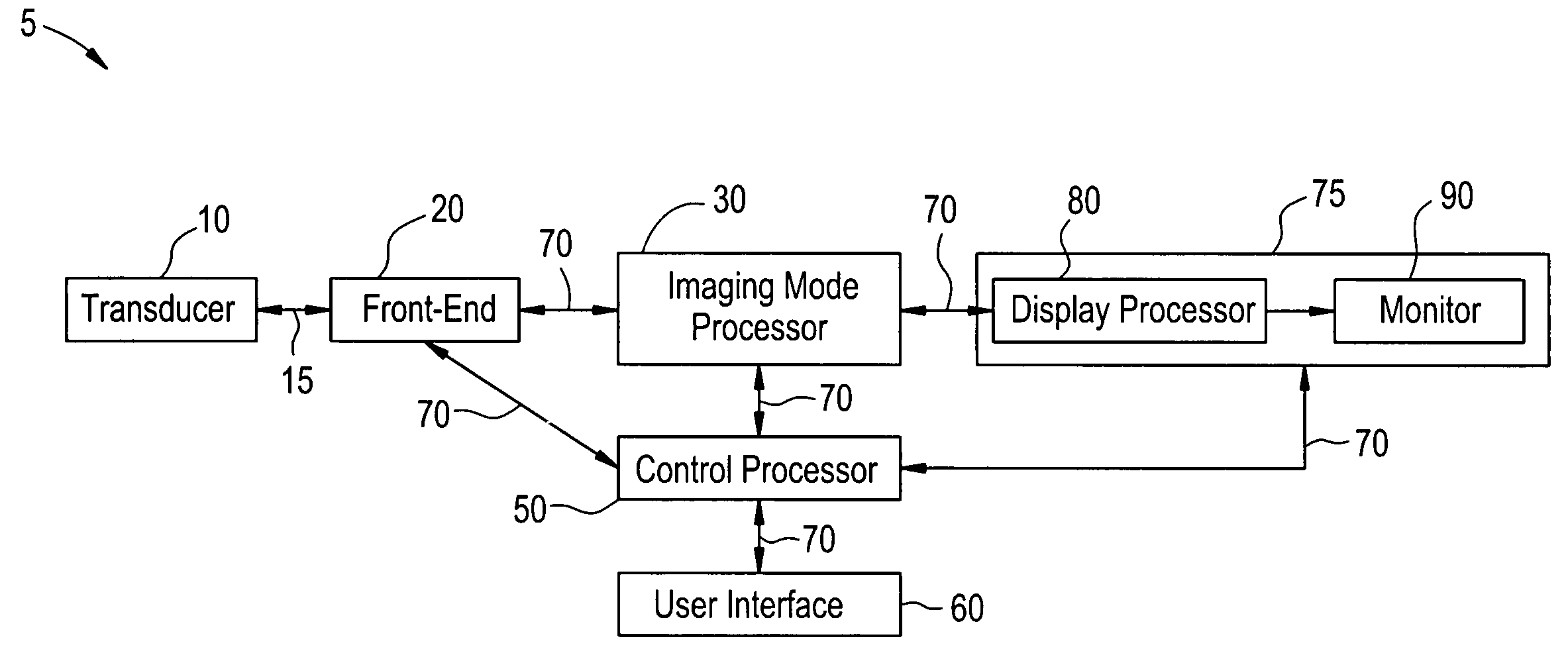
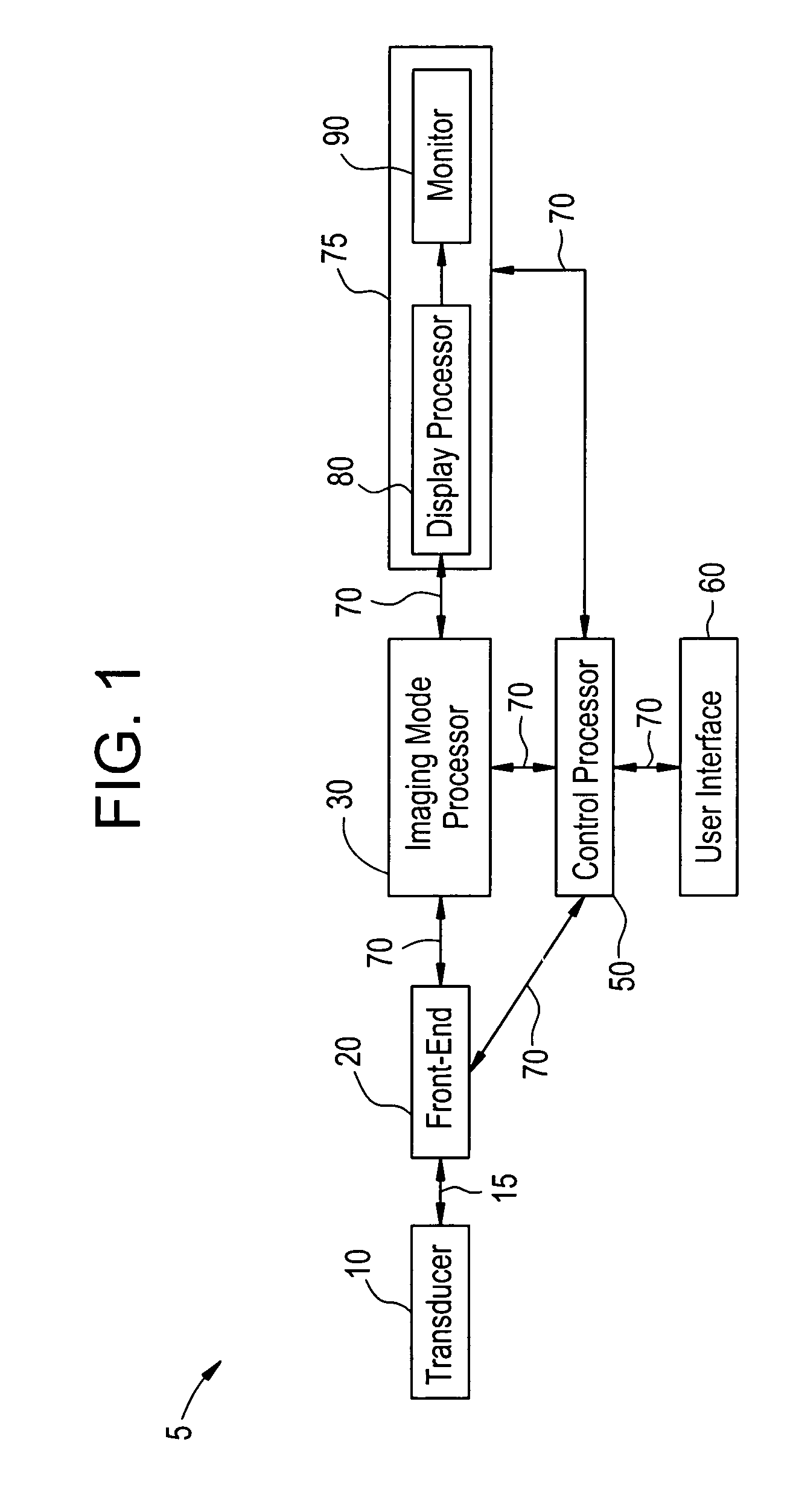
[0024]FIG. 1 illustrates a block diagram of an ultrasound imaging system 5 used in accordance with an embodiment of the present invention. The system 5 includes a transducer 10, a front-end 20, an imaging mode processor 30, a user interface 60, a control processor 50, and a display 75. The imaging mode processor 30 and the control processor 50 may be part of a back-end system. The transducer 10 is used to transmit ultrasound waves into a subject by converting electrical analog signals to ultrasonic energy. The transducer 10 also is used to receive ultrasound waves that are backscattered from the subject by converting ultrasonic energy to analog electrical signals. The front-end 20 including a receiver, a transmitter, and a beamformer, is used to create transmitted waveforms, beam patterns, receiver filtering techniques, and demodulation schemes that are used for various imaging modes. The front-end 20 converts digital data to analog data and vice versa. The front-end 20 interfaces w...


PUM


Abstract

Certain embodiments of the present invention provide a method and system for improved ultrasound imaging using single transmission coded excitation. Certain embodiments include encoding a first ultrasound beam with a first code, transmitting the first ultrasound beam on a first path, encoding a second ultrasound beam with a second code, transmitting the second ultrasound beam on a second path and receiving echo signals from the first and second ultrasound beams. The codes may be complimentary Golay codes or other complimentary codes. The first and second paths may be adjacent scan lines. The method may also include match filtering the echo signals with corresponding matched filters. Match filtered echo signals along adjacent scan lines may be filtered, such as with a lateral averaging filter or other finite impulse response filter. Alternate complementary code transmission helps preserve or improve frame rate while maintaining signal-to-noise ratio improvement and range lobe cancellation.

Description

RELATED APPLICATIONS [0001] [Not Applicable]FEDERALLY SPONSORED RESEARCH OR DEVELOPMENT [0002] [Not Applicable]MICROFICHE / COPYRIGHT REFERENCE [0003] [Not Applicable]BACKGROUND OF THE INVENTION [0004] The present invention generally relates to ultrasound imaging. In particular, the present invention relates to single transmission of coded excitation signals for ultrasound imaging. [0005] Ultrasound is sound having a frequency that is higher than a normal person may hear. Ultrasound imaging utilizes ultrasound waves or vibrations in the frequency spectrum above normal human hearing, such as the 2.5-10 MHz range. Ultrasound imaging systems transmit ultrasound into a subject, such as a patient, in short bursts. Echoes are reflected back to the system from the subject. Diagnostic images may be produced from the echoes. Ultrasound imaging techniques are similar to those used in sonar and radar. [0006] A medical ultrasound system forms an image by sequentially acquiring echo signals from u...

Claims


Application Information

Patent Timeline
Patent Type & Authority Applications(United States)
IPC IPC(8): G01S7/52A61B8/00G01S15/89H04R3/00
CPCG01S7/52095G01S15/8959G01S7/52085G01S7/52046G01S7/52077G01S15/8961
Inventor HAO, XIAOHUICHIAO, RICHARD YUNGHAIDER, BRUNO
Owner GE MEDICAL SYST GLOBAL TECH CO LLC
Why Eureka
Try Eureka