Methods and compositions to reduce scattering of light during therapeutic and diagnostic imaging procedures

a technology for diagnostic imaging and scattering, applied in the field of in vivo imaging, can solve the problems of reducing the oxygen carrying ability of patients, severely restricting clinical imaging applications, etc., and achieves the effects of reducing scattering, reducing signal attenuation, and improving image quality

Inactive Publication Date: 2005-05-26
BOARD OF RGT THE UNIV OF TEXAS SYST
View PDF64 Cites 15 Cited by
  • Summary
  • Abstract
  • Description
  • Claims
  • Application Information

AI Technical Summary

Benefits of technology

[0013] The present invention solves such needs in the art by providing a range of improved imaging methods with important applications in imaging through blood-rich structures. The invention particularly provides improved light-based imaging and treatment methods, including OCT imaging, using low-scattering, oxygen-carrying blood substitutes, such as hemoglobin solutions. The surprising use of the invention significantly reduces signal attenuation due to scattering from blood without resorting to the limitations of saline flushes. The invention thus simultaneously reduces scattering and significantly improves image quality whilst maintaining tissue oxygenation, avoiding ischemia and related tissue damage. The methods, compositions, kits and uses of the invention are therefore ideal for rapid and high resolution imaging of cardiovascular, brain and other highly vascularized tissues.

Problems solved by technology

Whether using saline, dextran or another contrast agent, the inventors realized that the attendant reduction in oxygen carrying ability would severely limit any clinical imaging applications, and would essentially prevent imaging in organs and tissues that are largely intolerant to ischemia, such as the heart and brain.

Method used


Image

  • Methods and compositions to reduce scattering of light during therapeutic and diagnostic imaging procedures
  • Methods and compositions to reduce scattering of light during therapeutic and diagnostic imaging procedures

Examples


example 1

[0124] Low-Scattering, Oxygen-Carrying Blood Substitutes for Improved Imaging

[0125] The present example describes the criteria for designing or selecting low-scattering, oxygen-carrying blood substitutes for use in improved imaging and related light-based techniques, and the validation of the selection criteria in in vitro studies.

A. Methods

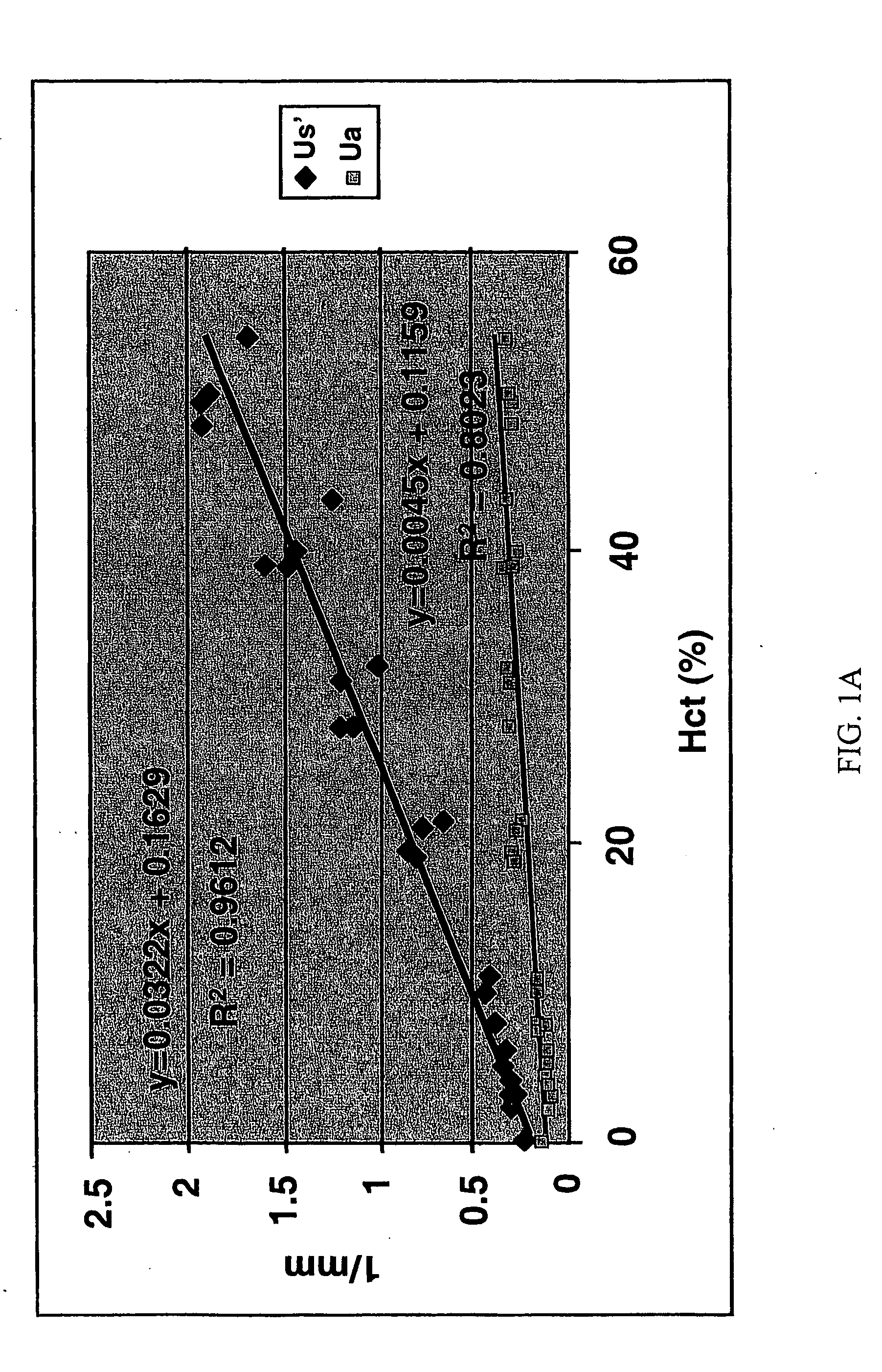
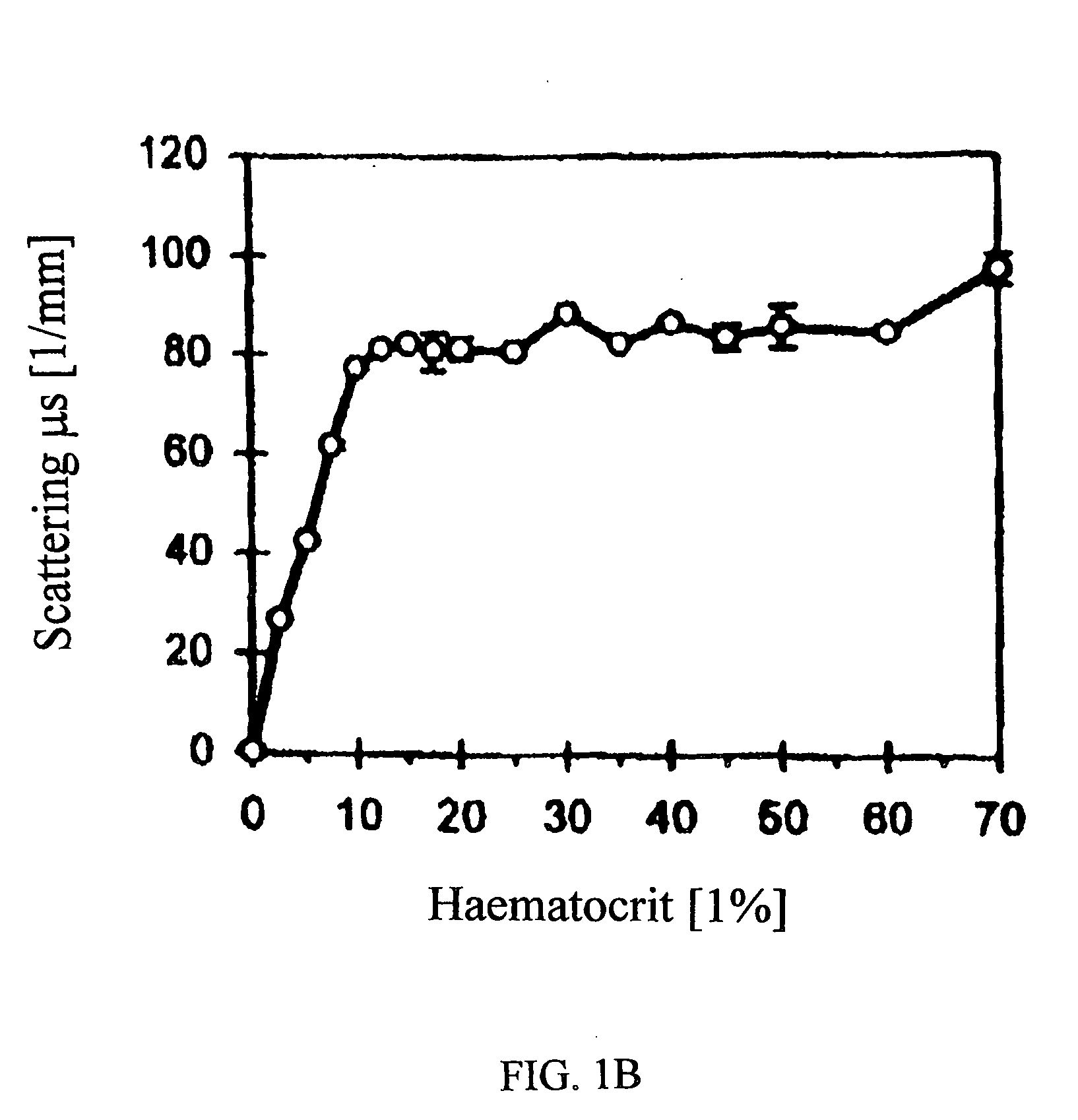
[0126] In vitro measurements were performed to determine the scattering and absorption properties of murine blood, blood substitute (Oxyglobin®), and dilutions of murine blood with Oxyglobin®. Oxyglobin® is isotonic to blood (prepared in a modified lactated Ringer's solution) and should not result in any hemolysis. An additional study was performed to specify the hematocrit reduction required for adequate visualization of the full RV thickness. For both studies, dilutions of fresh murine blood with Oxyglobin® (Biopure Corporation, Cambridge, Mass., www.biopure.com) were prepared to a hematocrit of 40, 30, 20, 10, 8, 5, and 3%. Pure blood (hem...

example 2

[0135] Improved OCT Myocardial Imaging with Low-Scattering Blood Substitutes

[0136] The present example in turns describes the successful in vivo imaging of cardiovascular tissues using low-scattering, oxygen-carrying blood substitutes prepared according to the selection criteria in Example 1.

A. Methods

1. OCT Instrumentation

[0137] The OCT system employed in the present example is based on a single-mode optical fiber Michelson interferometer and a semiconductor amplifier light source. The source has a center wavelength of 1310 nm and a spectral bandwidth of 60 nm. To visualize light incident on the murine myocardium with the unaided eye, a wavelength division multiplexer was used to combine 630 nm red light with 1310 nm source. A rapid scan optical delay line (Tearney et al., 1997) enables an image of a dynamic tissue, such as a beating murine heart, to be acquired at video rate. The axial and lateral resolutions were 16 μm and 12 μm, respectively. One A-scan (a depth scan) corr...


PUM


Abstract

Disclosed are improved methods and compositions for use in light-based in vivo imaging and treatment. The techniques described involve the use of low-scattering, oxygen-carrying blood substitutes in imaging and treatment methods, including OCT imaging. The invention has particular advantages in imaging within the cardiovascular system and highly vascularized or oxygen-dependent tissues.

Description

[0001] The present application claims priority to U.S. provisional application Ser. No. 60 / 348,604, filed Jan. 15, 2002, the entire text and figures of which are incorporated herein by reference without disclaimer.[0002] The U.S. Government owns rights in the present invention pursuant to grant numbers HL-59472 and HL-67475 from the National Institutes of Health.BACKGROUND OF THE INVENTION [0003] 1. Field of the Invention [0004] The present invention relates most substantially to the field of in vivo imaging. The invention provides methods and compositions for use in improved light-based imaging and treatment modalities based upon the use of low-scattering, oxygen-carrying blood substitutes. The use of hemoglobin solutions in combination with OCT imaging is a particular example within the invention. The compositions, methods and uses of the invention have important advantages in imaging tissues that are intolerant of ischemia. [0005] 2. Description of Related Art [0006] Non-invasive...

Claims


Application Information

Patent Timeline
Patent Type & Authority Applications(United States)
IPC IPC(8): A61B5/00
CPCA61B5/0066A61B5/0073A61B5/416A61B5/14551A61B5/14553A61B5/14535
Inventor VILLARD, JOSEPH W.FELDMAN, MARC D.MILNER, THOMAS E.
Owner BOARD OF RGT THE UNIV OF TEXAS SYST
Why Eureka
Try Eureka