
Device and method for facilitating hemostasis of a biopsy tract

a biopsy tract and hemostasis technology, applied in the field of wound closure devices, can solve the problems of small gauge needles, less satisfactory biopsy specimens, and difficulty in delivering sponges, and the technique has not reached widespread us

Inactive Publication Date: 2005-05-26
BOSTON SCI SCIMED INC
View PDF99 Cites 30 Cited by
  • Summary
  • Abstract
  • Description
  • Claims
  • Application Information

AI Technical Summary

Benefits of technology

The present invention is about a device and method for stopping bleeding from a biopsy or puncture wound in the breast. The device includes a catheter and an adaptor for delivering a hydrated sponge to the wound through a cannula. The sponge is injected into the tissue to stop bleeding and facilitate the healing process. The method involves removing tissue from the breast through the cannula and delivering the sponge to the wound to stop bleeding. The invention helps to improve the accuracy and safety of breast biopsy procedures.

Problems solved by technology

Possible complications of needle biopsy include bleeding at the biopsy site.
Small gauge needles, however, produce less satisfactory biopsy specimens but frequently are favored over larger bored needles because of their perceived safety.
Despite the advantages of using absorbable sponge to plug a biopsy tract this technique has not achieved widespread use because of difficulty in preparing and delivering the dry sponge material into the biopsy tract.
However, the sponge is difficult to deliver and expands slowly once delivered.
In addition, this delivery method can only deliver a sponge of a limited size which provides less local compression than desired and may incompletely fill the target site.
Further, bleeding may continue along sections of the biopsy tract where no sponge has been delivered.
However, the increased amount of tissue removed increases the potential for bleeding, hematoma, echimosis, and the like.
The permanently implanted clips can prove problematic, as they can migrate.

Method used


Image

  • Device and method for facilitating hemostasis of a biopsy tract
  • Device and method for facilitating hemostasis of a biopsy tract
  • Device and method for facilitating hemostasis of a biopsy tract

Examples


Embodiment Construction

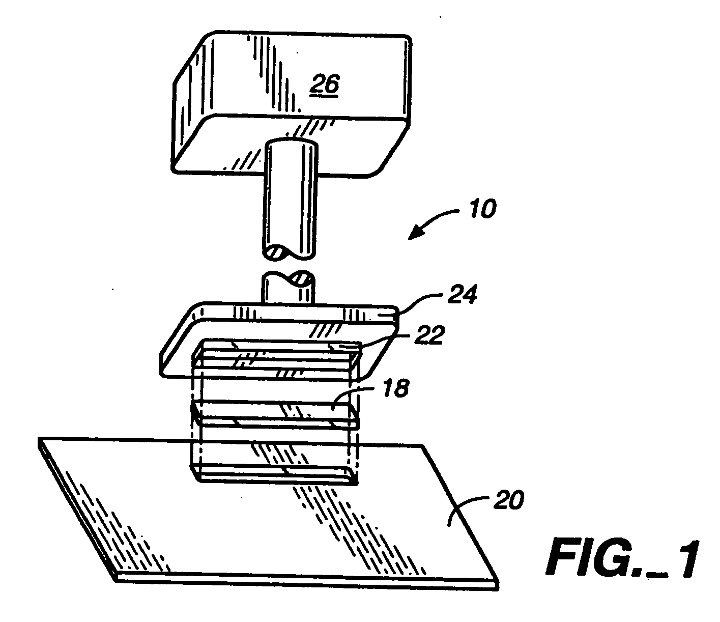
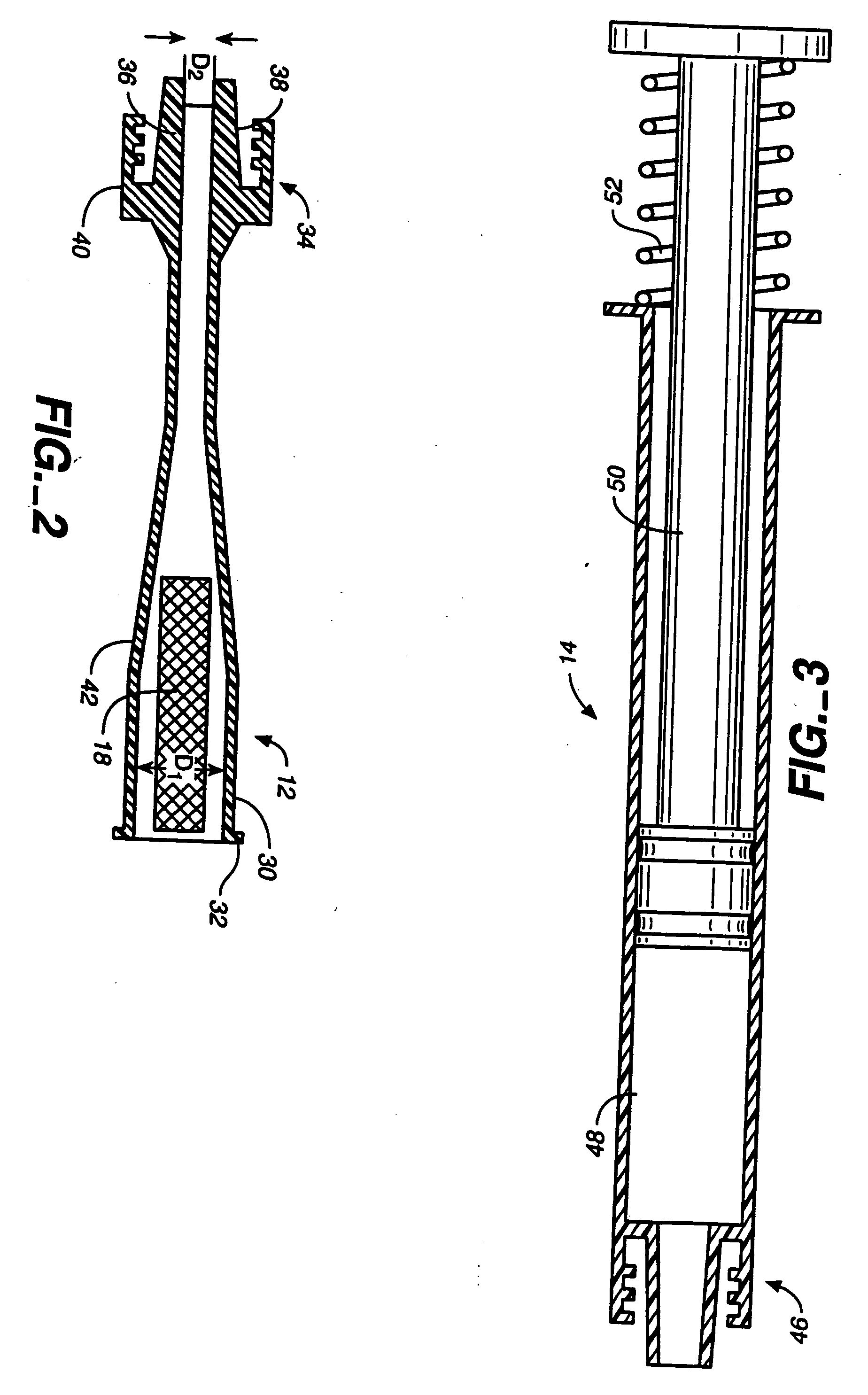
[0033] The system of the present invention delivers an absorbable sponge material in a hydrated state to facilitate hemostasis of a biopsy tract or other puncture wound in a simple and safe manner. The apparatus for delivering a hydrated absorbable sponge will be described below in connection with treatment of a biopsy tract after a percutaneous needle biopsy. However, the invention may be used for facilitating hemostasis of other types of puncture wounds or tissue access tracts to prevent bleeding of these wounds. The apparatus described with respect to FIGS. 1-7 is used for delivery of sponge material into all types of biopsy tracts in many different organs and tissues. The apparatus described with respect to FIGS. 8-12 is particularly designed for delivery of sponge material after biopsy with a breast biopsy device commonly known as a mammatome, however, this system can also be used for treatment of other biopsy sites and other types of wounds.

[0034] The system for facilitating ...


PUM


Abstract

A biopsy cannula and a delivery catheter are configured to deliver one or more absorbable sponge pledgets to a biopsy site after removal of one or more tissue samples from the site. The delivery catheter allows a large amount of hydrated sponge material to be delivery to the biopsy site to facilitate hemostasis. One example of the delivery catheter includes a closed distal end, a side port, a tapered section, and an enlarged proximal portion for receiving the pledget. The side port of the delivery catheter is arranged to delivery the pledget through the side port of the biopsy cannula. In order to fill a relatively large biopsy site where multiple tissue samples have been taken in a radial pattern, the biopsy cannula is rotated and additional pledgets are delivered to the biopsy site at different radial locations. The absorbable sponge pledget may also be used as a marker for location of the biopsy site at a later time.

Description

[0001] This application is a continuation of U.S. application Ser. No. 09 / 382,160, filed Aug. 24, 1999, which is a continuation-in-part of U.S. application Ser. No. 09 / 247,880, filed Feb. 10, 1999, which is a continuation-in-part of U.S. application Ser. No. 09 / 071,670, filed May 1, 1998, each of which is incorporated herein by reference in its entirety.BACKGROUND OF THE INVENTION [0002] 1. Field of the Invention [0003] The invention relates to a wound closure device, and more particularly, the invention relates to a device and method for facilitating hemostasis of a biopsy site or other puncture wound by injection of an absorbable sponge. [0004] 2. Brief Description of the Related Art [0005] Percutaneous needle biopsy of solid organs is one of the most common interventional medical procedures. Millions of percutaneous needle biopsies are performed annually in the United States and throughout the world. Percutaneous biopsy is a safe procedure which has supplanted surgical biopsy for...

Claims


Application Information

Patent Timeline
Patent Type & Authority Applications(United States)
IPC IPC(8): A61B10/00A61B10/02A61B17/00A61B17/03A61B17/04A61B17/08A61B19/00A61F2/00A61M27/00
CPCA61B10/02A61B10/0233A61B17/0057A61B2017/00654A61B2017/00004A61B2017/00637A61B19/54A61B90/39
Inventor ASHBY, MARKWHALEN, VAUGHN P.SING, EDUARDO CHI
Owner BOSTON SCI SCIMED INC
Learn More