Eureka AIR delivers breakthrough ideas for toughest innovation challenges, trusted by R&D personnel around the world.

Method for producing result images for an examination object

Inactive Publication Date: 2005-07-07
SIEMENS AG
View PDF14 Cites 45 Cited by
  • Summary
  • Abstract
  • Description
  • Claims
  • Application Information

AI Technical Summary

Benefits of technology

[0009] It is therefore an object of an embodiment of the present invention to provide an alternative method and an image processing system for automatically producing result images for the examination object using previously produced section image data, which allow diagnoses—particularly intermediate diagnoses for continuing the examination—to be produced significantly more easily, more quickly and more safely.

Problems solved by technology

Producing an intermediate diagnosis of this type requires a time involvement which is not to be underestimated, and this impairs the entire examination workflow.
A further problem is that identifying pathologies of particular internal structures, particularly in the case of very complex anatomical structures, in the section image data can be extremely difficult and requires some experience.
This may sometimes result in impairment of the quality of the section image examinations.
However, all of these methods only ever involve the output of just one model, and any diagnosis or intervention planning which is based on this is accordingly highly dependent on the quality of the model produced.

Method used


Image

  • Method for producing result images for an examination object
  • Method for producing result images for an examination object
  • Method for producing result images for an examination object

Examples


Embodiment Construction

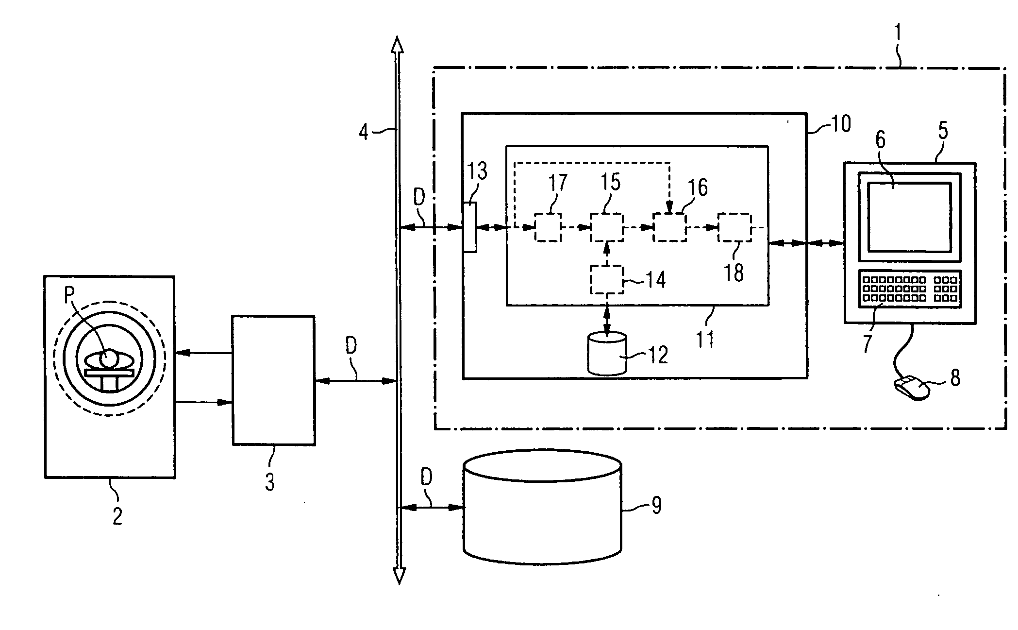
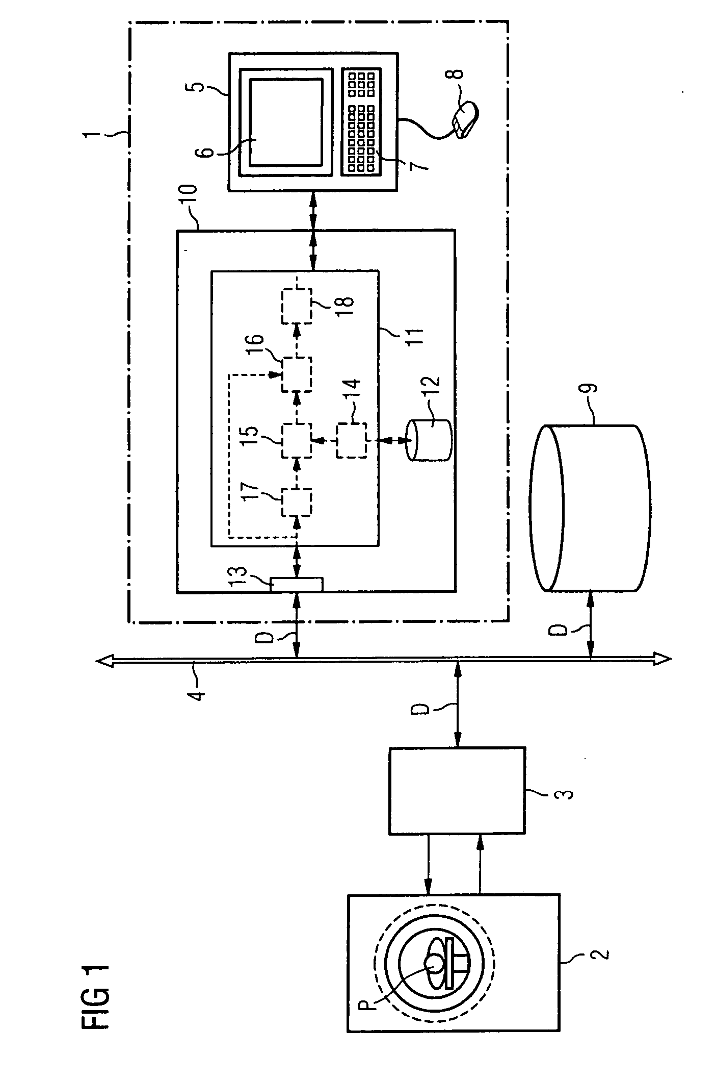
[0067] The exemplary embodiment of an inventive image processing system 1 which is shown in FIG. 1 essentially includes an image computer 10 and a console 5, connected thereto, or the like with a screen 6, a keyboard 7 and a pointer device 8, in this case a mouse 8. This console 5 or another user interface may also be used, by way of example, by the user to input the diagnostic questionnaire or to select it from a database containing prescribed diagnostic questionnaires.

[0068] The image computer 10 may be a computer of ordinary design, for example a workstation or the like, which may also be used for other image evaluation operations and / or to control image recorders (modalities) such as computer tomographs, magnetic resonance tomographs, ultrasound equipment etc. Fundamental components within this image computer 10 are, inter alia, a processor 11 and an interface 13 for receiving section image data D from a patient P which have been measured by a modality 2, in this case a magneti...


PUM


Abstract

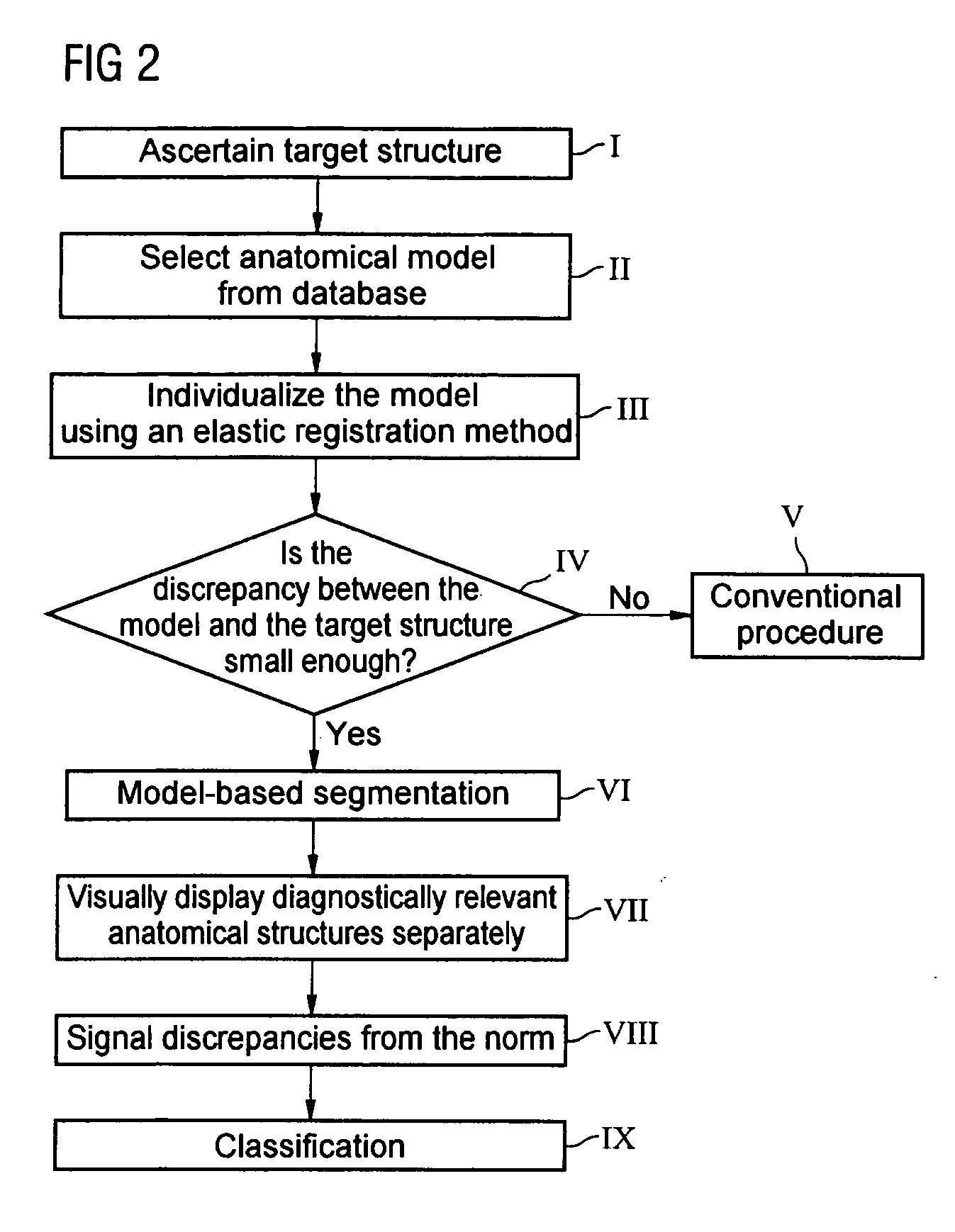
A method is for automatically producing result images for an examination object using section image data. In this case, a target structure is first of all ascertained in the section image data on the basis of a diagnostic questionnaire, and the target structure is taken as a basis for selecting an anatomical norm model whose geometry can be varied using model parameters. The norm model is automatically adapted to the target structure. The section image data are then segmented on the basis of the adapted norm model, with anatomical structures of the examination object which are relevant to the diagnostic questionnaire being separated by selecting all of the pixels within the section image data which are situated within a contour of the adapted norm model and / or at least one model part in line with the relevant structures or have a maximum discrepancy therefrom by a particular value. The relevant structures are then visually displayed separately and / or are stored for later visual display. The document also describes a corresponding image processing system.

Description

[0001] The present application hereby claims priority under 35 U.S.C. §119 on German patent application number DE 103 57 205.8 filed Dec. 8, 2003, the entire contents of which are hereby incorporated herein by reference. FIELD OF THE INVENTION [0002] The invention generally relates to a method for automatically producing result images for an examination object using section image data from the examination object in question. The invention also generally relates to an image processing system which can be used to carry out such a method. BACKGROUND OF THE INVENTION [0003] The result of examinations using modalities which produce section images, such as computer tomographs, magnetic resonance tomographs and ultrasound equipment, normally includes a number of series of section images of the examination object in question. For further planning of the examination and / or in order to produce a diagnosis, these section image data must in many cases be processed further during the examination...

Claims


Application Information

Patent Timeline
IPC IPC(8): A61B6/00A61B5/05A61B5/055A61B6/02A61B6/03A61B8/00A61B8/13A61B19/00G06F17/00G06F19/00G06T1/00G06T5/00G06T7/00G06T15/08
CPCA61B5/055A61B5/4504A61B5/4528A61B6/032A61B8/00G06T2210/41G06T7/0083G06T7/0089G06T19/00G06T2207/10081G06T2207/30008G06T7/0012G06T7/12G06T7/149A61B6/00
Inventor TANK, MARTIN
Owner SIEMENS AG
Why Eureka
Eureka Blog
Learn More